Abstract: Objective: To evaluate the effects of stannous fluoride (SnF2) toothpaste formulations, including one containing hops beta (β)-acids extract, on extracellular DNA (eDNA) within the extracellular polymeric substance (EPS) matrix of human saliva–derived in vitro oral biofilms. Methods: Disruption was evaluated using DNA-specific antibodies and high-throughput microarray polymer profiling, with disruption detected by reduction in antibody binding to microarrays (epitope depletion). The impact of different toothpaste formulations, including one with hops β-acids, was analyzed using scanning electron microscopy (SEM) imaging. In vitro–derived biofilms were cultured from pooled human saliva (from healthy volunteers) and printed onto nitrocellulose microarrays. Epitope depletion was used to assess the impact of five different SnF2 toothpaste compositions, including one containing hops β-acids, and a sodium monofluorophosphate toothpaste on the overall abundance and density of oral biofilm DNA within the EPS matrix of in vitro oral biofilms. Results: A toothpaste composition containing SnF2-gluconate/citrate-hops (0.454% SnF2) impacted both z-form DNA (z-DNA) and single-stranded DNA (ss-DNA) epitopes in oral biofilm EPS. Specifically, this composition elicited the greatest reduction in z-DNA present in oral biofilm EPS across all formulations tested (P < 0.05, aerobic and anaerobic biofilms). This treatment composition also visually reduced the abundance and density of the EPS matrix. Conclusions: Addition of hops β-acids drives the selective depletion of z-DNA epitopes, consistent with a model in which hops-containing formulations modify the EPS matrix through physicochemical interactions that impact specific structural domains, thereby contributing to changes in biofilm integrity and organization, observed through SEM. Clinical Implications: These findings provide a molecular-level rationale for formulation strategies that disrupt the EPS of adherent oral biofilms through surfactant-based modification and an experimental framework to determine which components of the EPS are most disrupted.
Keywords: hops, oral plaque, microarray polymer profiling, toothpaste, oral hygiene
Oral biofilms consist of microorganisms embedded in a sticky 3-dimensional matrix of extracellular polymeric substances (EPS), along with accumulated salivary proteins, minerals, and food debris that can lead to malodor.1,2 The EPS matrix, comprising a heterogenous mixture of extracellular DNA (eDNA), polysaccharides, proteins, and other macromolecules, governs the emergent properties of biofilms, including surface adhesion, mechanical cohesion, and resistance to removal.3,4 In the oral environment, EPS plays a defining role in the structure of oral biofilms and the need for daily hygiene by facilitating biomass attachment to surfaces and accumulation of unwanted byproducts through the fostering of a hydrated viscoelastic structure that resists physical disruption and active penetration.5,6
Within adherent oral biofilms, EPS is not a uniform material but rather a functionally differentiated composite. Extracellular polysaccharides, such as glucans and dextrans, synthesized primarily by oral Streptococci, form adhesive scaffolds that promote surface colonization and intercellular binding.7 Other polymers, including eDNA, have emerged as ubiquitous and critical structural components of biofilms, contributing to both early-stage adhesion and stabilization of the developing matrix.8 These distinct yet interacting components underpin the architecture of adherent oral biofilms as a cohesive, 3-dimensional system.9
eDNA has been recognized as a multifunctional contributor to biofilm formation and stability.10 Derived from cellular lysis or active secretion, eDNA participates in initial bacterial adhesion to surfaces, promotes aggregation of biomass, and contributes to the formation of a polymeric network within the EPS matrix through interactions with polysaccharides and proteins derived from bacterial or human cells.11 Structural analyses further indicate that eDNA forms extended networks that reinforce biofilm cohesion and maintain integrity under applied stress, such as toothbrushing.8
Despite advances in characterizing EPS composition, the molecular-level organization and heterogeneity of its constituent components remain poorly understood. In particular, eDNA has largely been treated as a homogenous structural polymer, with limited resolution of its potential conformational or context with the “matrixome,” in which distinct components contribute differently to biofilm architecture and function.6,11 Resolving such heterogeneity is essential to understanding how biofilm structure can be modified or destabilized by external agents.
Molecular probes and microarray polymer profiling (MAPP) are powerful analytical tools used extensively for high-throughput polymer analysis in natural and industrial systems.12-19 MAPP exploits the specificity of molecular probes to decipher complex mixtures of molecular components within extracted samples printed as microarrays. The capacity of enzymes (or other substances) to degrade or remove specific molecular components can be assessed by reduction in probe binding, an approach known as epitope depletion.20
This approach enables semi-quantitative assessment of changes in specific EPS components within complex adherent oral biofilms. The current study has leveraged MAPP and epitope depletion to assess and understand the impact of different toothpaste formulations, including one hops beta (β)-acids-containing paste, on specific eDNA structures within oral biofilm EPS (Figure 1).
Evidence presented in this special issue and elsewhere indicates that hops β-acids extract contributes to the physical modification of adherent oral biofilms, promoting dispersion and thinning of EPS-rich structures in both in vivo and in vitro systems.21,22 These findings support the role of hops β-acids extract as a surfactant that modifies the structural integrity of the biofilm/EPS matrix to facilitate its disruption and removal during toothbrushing.
In the present study, the authors investigated the impact of various stannous fluoride (SnF2) toothpaste formulations with different cleansing ingredients, including hops β-acids, and a sodium monofluorophosphate (MFP) toothpaste on changes in the eDNA component of oral plaque–derived EPS. This research complements associated work within this special issue investigating changes in biofilm structure, EPS distribution, and crosslinking ion content in different biofilms.22 Specifically, this study analyzed changes in z-form DNA (z-DNA) and single-stranded DNA (ss-DNA) (as assessed by changes in antibody binding) in EPS samples, following exposure to toothpaste formulations containing SnF2, with an additional formulation containing SnF2 and hops β-acids. By resolving these components, this work aims to define how structurally differentiated domains within eDNA contribute to EPS architecture and how they respond to treatment.
Methods and Materials
Oral Biofilms Growth and Treatment
Biofilms were grown from pooled oral lavage samples collected from at least eight healthy donors. Lavage was mixed with fresh media containing 3% tryptic soy broth and 1% yeast extract, supplemented with 0.005 mg/ml hemin and 0.0005 mg/ml menadione. After growing the culture 24 hours at 37°C in anaerobic conditions, the bacterial culture was used to inoculate 3D-printed discs in 24-well plates, with daily media replenishment over a 5-day growth period. On day 6, biofilms underwent a single 2-minute treatment with 1:3 toothpaste supernatant, followed by a 30-second water rinse, and were immediately fixed.
Sample Fixation for Scanning Electron Microscopy Visualization
Scanning electron microscopy (SEM) samples were fixed using a 2% glutaraldehyde solution in 1M phosphate buffer, pH 7.2. Tissues were submerged in 2 mL of the fixative per well (for 24-well plates) for 2 hours at room temperature, followed by overnight incubation at 4°C. Post-fixation, samples underwent an ethanol dehydration series: 50% and 70% ethanol (EtOH) for 45 minutes each at -20°C, then 85% EtOH for 30 minutes at 4°C, and 95% EtOH overnight at 4°C for long-term storage, culminating in two 100% EtOH steps before further processing. After dehydration, samples were mounted on a conductive disc using copper tape, posts trimmed down and sputter coated with a 15 µm layer of gold.
Microarray Polymer Profiling With Oral Biofilms
In vitro–derived biofilms were cultured from human saliva that was obtained from adult donors, who refrained from eating or drinking at least 2 hours prior to saliva collection. Artificial saliva growth medium was prepared by dissolving 0.20% (w/v) yeast extract, 0.50% (w/v) proteose peptone, 0.25% (w/v) type III hog gastric mucin, 0.02% (w/v) sodium chloride, 0.02% (w/v) potassium chloride, and 0.03% (w/v) calcium chloride in distilled water. The solution was sterilized and supplemented with 0.05% (w/v) urea. Biofilms were cultivated in sterile six-well tissue culture plates, under aerobic or anaerobic (in a Whitley A35 anaerobic chamber: 80% nitrogen, 10% carbon dioxide, 10% hydrogen) conditions, as indicated. Plates were incubated at 37°C for 48 hours (culture medium exchanged after 24 hours) for biofilm growth before biofilms were harvested and sonicated in 50 mM CDTA pH 7.5 for 10 minutes. Samples were incubated in a vertical tube rotator (Qiagen GmbH, qiagen.com) for 2 hours at an agitation setting of 10s-1, followed by EPS extraction with 4 M sodium hydroxide (NaOH) + 0.1% w/v sodium borohydride (NaBH4), for 2 hours at 10s-1. Extracted EPS was printed onto nitrocellulose microarrays using a non-contact microarray robot (Marathon, ArrayJet, arrayjet.com).23
Epitope Depletion With Toothpastes
MFP: 0.76% toothpaste (Colgate Cavity Protection, Colgate Palmolive, colgate.com)
SnF2-pyrophosphate-arginine: 0.454% SnF2 toothpaste (Colgate Total Plaque Pro Release, Colgate Palmolive)
SnF2-potassium nitrate (KNO3)/pyrophosphate: 0.454% SnF2 toothpaste (Colgate Total Active Prevention, Colgate Palmolive)
SnF2-tripolyphosphate: 0.454% SnF2 toothpaste (Parodontax Complete Protection, Haleon, parodontax.com)
SnF2-gluconate/citrate: 0.454% SnF2 toothpaste (Crest Pro-Health Advanced Bacteria Protection, The Procter & Gamble Company, crest.com)
SnF2-gluconate/citrate-hops: 0.454% SnF2 toothpaste with 0.15% hops β-acids (Crest Gum Detoxify with Activ Bio-Cleanser™, The Procter & Gamble Company)
Toothpaste slurries were prepared by diluting products of interest in water at a 1:9 (w/v) ratio. The mixtures were homogenized using a tissue lyser to ensure complete emulsification. Printed microarrays were incubated with these toothpaste slurries at ambient room temperature for 20 minutes, with slight agitation, followed by two 5-minute rinse steps in water to remove residual toothpaste, and subsequently washed overnight in phosphate-buffered saline (PBS).
Molecular Probing
Printed microarrays were blocked for 1 hour at room temperature in PBS supplemented with 5% (w/v) dried skimmed milk powder (Marvel Original, Batch 3025307, Premier Foods, premierfoods.co.uk). Following blocking, arrays were incubated for 2 hours with antibodies specific to z-DNA (Z22, Absolute Antibody, absoluteantibody.com) or ss/ds-DNA (A52, Absolute Antibody) in PBS supplemented with 5% (w/v) milk powder. Arrays were then washed thoroughly with PBS and incubated for 2 hours with alkaline phosphatase–conjugated secondary antibody (mouse, Category # 115-055-003, Jackson ImmunoResearch, jacksonimmuno.com) diluted 1 in 1,000 (v/v) in PBS containing 5% (w/v) milk powder. After being washed extensively in PBS, arrays were developed using 5-bromo-4-chloro-3-indolyl phosphate p-toluidine salt (BCIP, Merck, emdgroup.com) and nitro blue tetrazolium chloride (NBT, Melford Laboratories Ltd., melford.co.uk). Developed arrays were scanned, and the binding intensities of probes to individual spots were quantified using Array-Pro Analyzer software (Version 6.3). For heat map generation, the highest mean spot signal intensity was set to 100, and all other values were normalized accordingly.23 The values in Table 1 correspond to the mean of three technical replicates.
Statistical Analysis
To assess differences between treatment means, analysis of variance (ANOVA) was performed at a significance level (α) of 0.05 using JMP18.0. Statistically distinct groups were identified by post-hoc analysis (Tukey’s Honestly Significant Difference test) and displayed as letters derived from a connecting letters report. Treatments sharing a common letter were not significantly different at 95% confidence level. Groups that did not share any letters were significantly different.
Results
Figure 2a shows the SEM image of an oral biofilm treated with an MFP-containing toothpaste, revealing a notably dense and robust EPS matrix characterized by a tightly packed architecture and a lack of discernible spaces. In contrast, Figure 2b depicts an oral biofilm treated with an SnF2 toothpaste containing hops β-acids. The hops-treated EPS matrix exhibited pronounced physical disruption, with the emergence of numerous channels and visible structural compromises, indicating that hops β-acids weaken the biofilm by directly dispersing or altering the EPS layer, compromising its structural strength and making it more susceptible to further disruption.
A molecular probe that recognizes microbial z-DNA14 was used to assess the relative abundance of z-DNA present in cultured EPS, following treatment with different toothpaste formulations. Colgate Cavity Protection and Colgate Total Active Prevention toothpastes did not appear to significantly impact the epitope signal associated with z-DNA presented in aerobic biofilm, versus no treatment (Table 1). Parodontax and Colgate Total Plaque Pro Release toothpastes reduced the epitope signal associated with microbial z-DNA from 73.0% of the maximum detected z-DNA signal on the array to 52.3% and 57.7%, respectively. Crest Pro-Health Advanced containing gluconate/citrate SnF2 did not significantly reduce the relative signal of detectable z-DNA epitope versus no treatment (Table 1). Treatment with Crest Pro-Health Advanced in fact appeared to drive a slight increase from 73.0% of maximum detected signal (no treatment) to 96.8% of maximum detected signal, potentially suggesting epitope unmasking due to contained chemistry impacting other EPS components that interact with z-DNA. However, when another gluconate/citrate SnF2 toothpaste containing hops β-acids (Crest Gum Detoxify with Activ Bio-Cleanser) was used to treat the oral biofilms, there was a significant reduction in the detection of z-DNA epitope from 73.0% (no treatment) to 37.0% (Table 1).
Similar trends were observed for performance of the respective toothpastes against z-DNA present in anaerobic biofilm (Table 1). Colgate Cavity Protection (-6.3% vs no treatment) and Colgate Total Active Prevention (-0.8% vs no treatment) did not significantly increase or decrease the epitope signal associated with z-DNA presented in these biofilms versus no treatment. Parodontax (-23.8%) and Colgate Total Plaque Pro Release (-18.1%) both drove discernible reductions versus the no treatment control group, while the addition of hops β-acids extract to gluconate/citrate SnF2 (Crest Gum Detoxify) resulted in a 29.6% reduction in epitope signal associated with z-DNA versus no treatment. When benchmarked against another gluconate/citrate SnF2 toothpaste not containing hops (Crest ProHealth Advanced), there was a difference of 48.1% in detected epitope, indicating a marked difference in z-DNA depletion relative to the SnF2-only formulation against microbial soils produced by bacteria that thrive in anaerobic conditions.
A commercially available antibody (MAB3034) with reported 90% reactivity to single-stranded (ss)-DNA and 10% reactivity to double-stranded (ds)-DNA was also used to provide an indication of toothpaste efficacy against other configurations of DNA present in adherent oral biofilm–derived EPS. Interestingly, while Colgate Cavity Protection delivered a significant increase in the epitope signal associated with ss-DNA in both types of oral biofilms tested in this study versus no treatment, all other toothpastes tested delivered a significant reduction on the relative abundance of detectable ss-DNA epitope in aerobic or anaerobic biofilm, respectively (Table 1). Parodontax (29.6% and 32.2%), Colgate Total Active Prevention (42.6% and 54.8%), Colgate Total Plaque Pro Release (12.4% and 10.7%), Crest Pro-Health Advanced (7.7% and 8.4%), and Crest Gum Detoxify containing hops β-acids (13.1% and 11.1%) all significantly reduced the relative detection of ss-DNA, compared to no treatment in different biofilm types (78% and 81% of maximal ss-DNA signal, respectively).
Discussion
Hops β-acids extract functions as a plant-derived surfactant, contributing to interfacial modification within oral biofilm and potentially altering its structure. As a toothpaste formulation component, hops β-acids support physicochemical cleaning processes. A separate article in this special issue describes how hops-containing treatments reduce residual biofilm coverage and thickness compared to control treatments across multiple experiments and reduce EPS content as well as EPS normalized to bacteria biomass.22 These findings indicate selectivity toward the EPS contribution to the biofilm.
The present study demonstrates formulation-dependent differences in the impact of oral care products on extracellular eDNA epitopes within in vitro oral biofilm EPS. Among the formulations tested, the formulation containing hops β-acids extract in combination with SnF2 (Crest Gum Detoxify with Activ Bio-Cleanser) produced the most consistent effect with strongly detectable reduction in both z-DNA and ss-DNA epitopes, distinguishing its effect from both stannous-only and comparator formulations versus no treatment. In contrast, other products exhibited either minimal or inconsistent changes in detectable z-DNA and ss-DNA, highlighting that different chemistries interact with EPS components in distinct ways that are still to be determined. Nevertheless, this pattern is consistent with the broader observation that biofilm matrix composition and response to treatment are strongly dependent on formulation-specific physicochemical interactions between the EPS matrix and the treatment.
Interpretations of these findings must be considered within the framework of epitope depletion reinforced with SEM observations of EPS removal. Reductions in probe binding intensity reflect depletion of accessible epitopes and are qualitatively supported by the microscopy images showing breakdown and clear openings of the biofilm architecture.20 As such, observed decreases in detectable z-DNA and ss-DNA suggest reduction in EPS consistent with removal of eDNA. However, removal was not consistently observed between z-DNA and ss-DNA in response to all the tested formulations.
The differential behavior of z-DNA relative to more canonical ss/ds-DNA epitopes reflects the heterogeneity of eDNA within EPS,24 along with their varying susceptibilities to toothpaste formulations, demonstrated in the current study. Extracellular DNA is increasingly recognized as a critical architectural component of biofilms, contributing to both early-stage adhesion and matrix cohesion through the formation of extended polymeric networks and interactions with other EPS constituents.5 The present data extend this understanding by indicating that within the broader eDNA pool, z-DNA exhibits different susceptibilities to external disruption by toothpaste formulations. z-DNA tends to be stabilized by proteins or positively charged compounds such as spermidine25; it is possible that hops β-acids are depleting z-DNA through interactions with positively charged compounds that stabilize the rigid conformation.
In contrast, the more generally observed reductions in ss-DNA epitopes across multiple formulations are consistent with a general impact on eDNA. Previous studies have shown that eDNA participates in biofilm cohesion through distributed polymeric interactions that stabilize the matrix and promote aggregation.8,10 The relatively non-selective depletion of ss-DNA epitopes observed here suggests that many formulations can disrupt this general network to some extent, whereas the more pronounced and selective depletion of z-DNA with hops-containing formulations points toward preferential modification of specific structural domains within the EPS.
Future work integrating molecular probing using an expanded set of probes (against other EPS composites in oral plaque) together with imaging techniques will be required to resolve the localization and functional role of distinct DNA domains within intact biofilms. These insights will help further elucidate how formulation-driven modifications of EPS translate into enhanced biofilm removal and performance in oral care systems. Experiments such as these will help elucidate the role that the molecular “adhesive glues” play in adherent biofilm adhesion and cohesion, as well as validate strategies designed to disrupt them.
Conclusion
In the context of this special issue, which demonstrates that hops β-acids extract promotes biofilm thinning in solution when incorporated into toothpaste formulations, the present study provides additional molecular-level insights into these effects. The selective depletion of z-DNA epitopes is consistent with a model in which hops-containing formulations modify the EPS matrix through physicochemical interactions that impact specific structural domains, thereby contributing to changes in biofilm integrity and organization. The complementary SEM images reinforce that the reduction in epitope signal is associated with EPS disruption and removal. These findings cannot confirm direct interaction with or targeting of eDNA but rather support a broader mechanism involving modification of the EPS environment in which DNA is embedded, whether targeted or not.
ACKNOWLEDGMENT
The authors thank UKRI Innovate UK and Procter & Gamble for funding KTP project 10121458; Richard Holliday, PhD, (NIHR, Newcastle University) for the provision of oral plaque material for the current study; Max Kelly for technical expertise; and Lisa Sagel for support in preparation of the manuscript.
DISCLOSURE
HCLY, AM-B, HR, NJL, SSJ, and RS are employees of The Procter & Gamble Company. AG-C, IL, CSF, NSJ, and WGTW are employed by Newcastle University and declare no conflict of interests.
ABOUT THE AUTHORS
Hamish C.L. Yau, PhD
The Procter & Gamble Company, Newcastle upon Tyne, UK
Alina Goldberg-Cavalleri, PhD
School of Natural and Environmental Sciences, Newcastle University, Newcastle upon Tyne, UK; School of Dental Sciences, Faculty of Medical Sciences, Newcastle University, Newcastle upon Tyne, UK; and NIHR Newcastle Biomedical Research Centre (BRC), Newcastle upon Tyne, UK
Alejandra Muñoz-Bodnar, PhD
The Procter & Gamble Company, Mason, Ohio
Heather Rice, AS
The Procter & Gamble Company, Mason, Ohio
Ieva Lelėnaitė, PhD
School of Natural and Environmental Sciences, Newcastle University, Newcastle upon Tyne, UK
Charlotte S. Fletcher, BSc
School of Natural and Environmental Sciences, Newcastle University, Newcastle upon Tyne, UK
Neil J. Lant, PhD
The Procter & Gamble Company, Newcastle upon Tyne, UK
Nicholas S. Jakubovics, PhD
School of Dental Sciences, Faculty of Medical Sciences, Newcastle University, Newcastle upon Tyne, UK; and NIHR Newcastle Biomedical Research Centre (BRC), Newcastle upon Tyne, UK
Samuel St. John, PhD
The Procter & Gamble Company, Mason, Ohio
Ross Strand, BS
The Procter & Gamble Company, Mason, Ohio
William G.T. Willats, PhD
School of Natural and Environmental Sciences, Newcastle University, Newcastle upon Tyne, UK
References
1. Tonzetich J. Production and origin of oral malodor: a review of mechanisms and methods of analysis. J Periodontol. 1977;48(1):13-20.
2. Koo H, Falsetta ML, Klein MI. The exopolysaccharide matrix: a virulence determinant of cariogenic biofilm. J Dent Res. 2013;92(12):1065-1073.
3. Flemming HC, Wingender J. The biofilm matrix. Nat Rev Microbiol. 2010;8(9):623-633.
4. Jakubovics NS, Goodman SD, Mashburn-Warren L, et al. The dental plaque biofilm matrix. Periodontol 2000. 2021;86(1):32-56.
5. Flemming HC, van Hullebusch ED, Little BJ, et al. Microbial extracellular polymeric substances in the environment, technology and medicine. Nat Rev Microbiol. 2025;23(2):87-105.
6. Karygianni L, Ren Z, Koo H, Thurnheer T. Biofilm matrixome: extracellular components in structured microbial communities. Trends Microbiol. 2020;28(8):668-681.
7. Flemming HC, van Hullebusch ED, Neu TR, et al. The biofilm matrix: multitasking in a shared space. Nat Rev Microbiol. 2023;21(2):70-86.
8. Sharma DK, Rajpurohit YS. Multitasking functions of bacterial extracellular DNA in biofilms. J Bacteriol. 2024;206(4):e0000624.
9. Koo H, Xiao J, Klein MI, Jeon JG. Exopolysaccharides produced by Streptococcus mutans glucosyltransferases modulate the establishment of microcolonies within multispecies biofilms. J Bacteriol. 2010;192(12):3024-3032.
10. Campoccia D, Montanaro L, Arciola CR. Extracellular DNA (eDNA). A major ubiquitous element of the bacterial biofilm architecture. Int J Mol Sci. 2021;22(16):9100.
11. Rath S, Fatma S, Das S. Unraveling the multifaceted role of extracellular DNA (eDNA) of biofilm in bacterial physiology, biofilm formation, and matrixome architecture. Crit Rev Biochem Mol Biol. 2025;60(1-3):1-32.
12. Isager Ahl L, Pedersen HL, Jørgensen B, et al. Exploring the polysaccharide composition of plant cell walls in succulent aloes. Plants, People, Planet. 2023;5(3):335-353.
13. Bakshani CR, Cuskin F, Lant NJ, et al. Analysis of glycans in a Burnt-on/Baked-on (BoBo) model food soil using microarray polymer profiling (MAPP) and immunofluorescence microscopy. Food Chem. 2023;410:135379.
14. Yau HCL, Malekpour AK, Momin NG, et al. Removal of eDNA from fabrics using a novel laundry DNase revealed using high-resolution imaging. Sci Rep. 2021;11(1):21542.
15. Yau HCL, Byard J, Thompson LE, et al. Enzymatic modification of cotton fibre polysaccharides as an enabler of sustainable laundry detergents. Sci Rep. 2024;14(1):22049.
16. Lant NJ, Momin NG, Morales Garcia A, et al. Methods for making cleaning compositions and detecting soils. US20220373467. 2022.
17. Kobel CM, Leu A, Vera-Ponce de León A, et al. Protozoal populations drive system-wide variation in the rumen microbiome. Nat Commun. 2025;16(1):6238.
18. Fangel JU, Sørensen KM, Jacobsen N, et al. The legacy of terrestrial plant evolution on cell wall fine structure. Plant Cell Environ. 2024;47(4):1238-1254.
19. Cesari AB, Castilla Marín VE, Muratore LN, et al. New inoculation strategy for legume based on rhizobium-metabolite co-encapsulation. In: Medina C, López-Baena FJ, eds. Host-Pathogen Interactions. Methods in Molecular Biology. Vol 2751. New York, NY: Humana Press; 2024.
20. Vidal-Melgosa S, Pedersen HL, Schückel J, et al. A new versatile microarray-based method for high throughput screening of carbohydrate-active enzymes. J Biol Chem. 2015;290(14):9020-9036.
21. Zsiska M, Anastasia MK, Baig AA, St. John S. Enhanced dental biofilm removal with toothpastes containing hops β-acids extract: in vivo studies on oral hygiene. Compend Contin Educ Dent. 2026;47 suppl 3:22-27.
22. St. John S, Strand R. Effects of hops β-acids extract on the structure and composition of mature and immature oral biofilms. Compend Contin Educ Dent. 2026;47 suppl 3:14-21.
23. Moller I, Marcus SE, Haeger A, et al. High-throughput screening of monoclonal antibodies against plant cell wall glycans by hierarchical clustering of their carbohydrate microarray binding profiles. Glycoconj J. 2008;25(1):37-48.
24. Buzzo JR, Devaraj A, Gloag ES, et al. Z-form extracellular DNA is a structural component of the bacterial biofilm matrix. Cell. 2021;184(23):5740-5758.e17.
25. More KR, Buzzo JR, Devaraj A, et al. Spermidine is a common component of the eDNA-dependent biofilm matrix. NPJ Biofilms Microbiomes. 2026;12(1):141.
Figures and Images
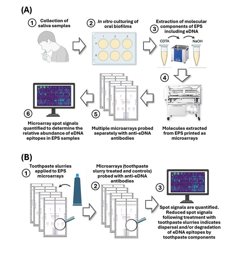
Figure 1
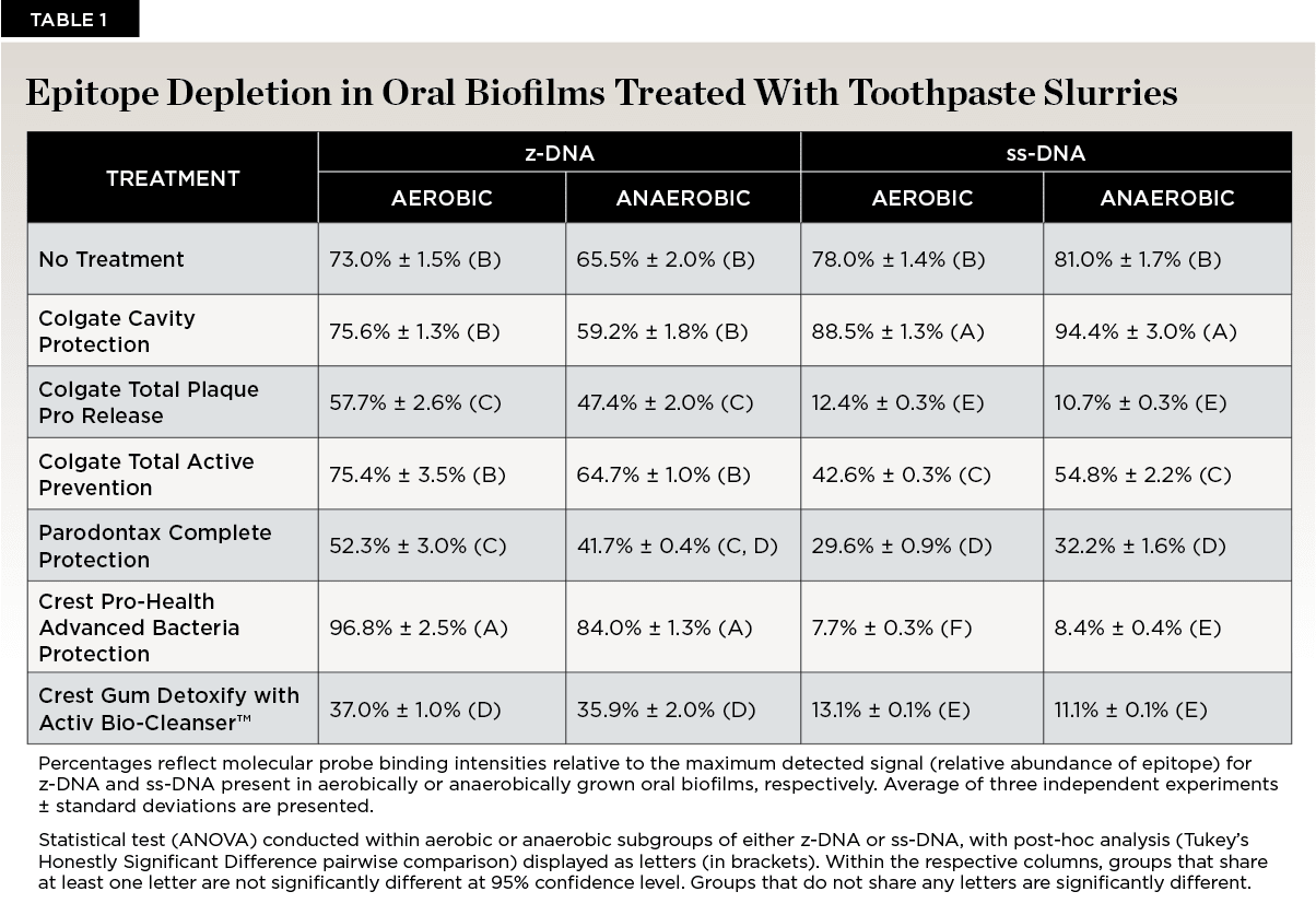
Table 1
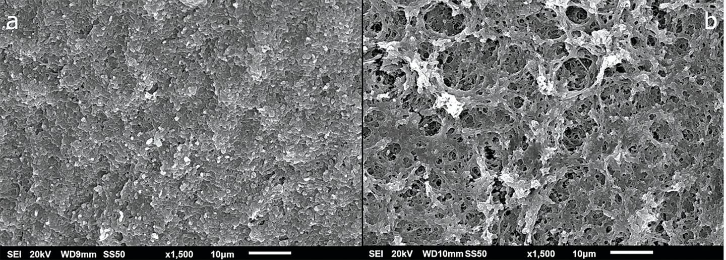
Figure 2
