Abstract: Background: The extracellular polymeric substance (EPS) matrix governs the cohesion, adhesion, and mechanical resilience of oral biofilms, limiting the effectiveness of physical removal during oral hygiene. Understanding how surfactant systems interact with EPS is a critical step in linking formulation-level surface activity to clinically measurable biofilm removal. Methods: In situ–derived oral biofilms were grown on artificial enamel (hydroxyapatite) discs that were mounted on a dental appliance worn by participants then treated ex situ with hops beta (β)-acids extract (Activ Bio-Cleanser™)–containing solutions or toothpastes versus surfactant (dishwashing liquid) and placebo controls. Biofilm structure, thickness, and composition were assessed using confocal laser scanning microscopy (CLSM) with fluorescent labeling of bacterial mass, EPS, and calcium. Complementary experiments evaluated the effects of hops on immature biofilms formed from salivary pellicle coatings. Results: Hops β-acids-containing solutions and toothpastes reduced biofilm coverage and thickness relative to comparator surfactant solutions and control toothpastes (P < 0.05). CLSM imaging demonstrated disruption and thinning of the EPS matrix, accompanied by reduced biofilm cohesion and adhesion to enamel surfaces. Hops-containing treatments also altered immature biofilm structure on artificial enamel, consistent with surfactant-driven physicochemical interactions at the tooth surface. Conclusions: The results demonstrate that hops β-acids extracts function as surfactant-based excipients capable of dispersing and thinning EPS-rich, in situ–derived oral biofilms following ex vivo treatment and analysis. Within the context of this special issue, this study provides a mechanistic link between fundamental surfactant behavior and subsequent human studies, supporting a formulation-focused framework in which EPS disruption enhances the efficiency of biofilm removal during toothbrushing. Practical Implications: These findings support the role of hops to enhance the efficacy of toothpaste formulations for biofilm removal, helping to improve oral hygiene efficiency for patients.
Keywords: surfactant, extracellular polymeric substances, hops β-acids, dental hygiene, toothpaste
Oral bacteria colonize tooth surfaces as structured biofilms whose cohesion and adhesion are governed by an extracellular polymeric substance (EPS) matrix.1,2 This EPS framework confers mechanical resilience and surface attachment, limiting the effectiveness of physical removal during oral hygiene.3-5 While toothbrushing remains the primary means of biofilm control, residual adherent biofilm commonly persists.5-9 Rather than relying solely on mechanical force, new formulation strategies that modify the physical properties of biofilm to reduce its cohesive and adhesive properties to facilitate its removal would be beneficial.">
In general, EPS promotes bacterial aggregation and surface attachment and protects bacteria from desiccation, predation, antimicrobial agents, antibodies, and bacteriophages.10,11 Likewise, the EPS matrix establishes diffusion gradients in the oral biofilms that encourage maturation of bacterial films into pathogenic forms.1,3,12-14 The structural stability promoted by EPS makes understanding its removal a key motivation for research. From a formulation perspective, surfactants offer a physicochemical means to interact with EPS and encourage biofilm dispersion during toothbrushing. However, despite their routine inclusion in toothpastes for foaming and flavor emulsification, surfactant systems have historically been under-investigated for their ability to directly influence EPS integrity and biofilm cohesion. Establishing how surfactant-based excipients interact with EPS-rich oral biofilms is therefore a critical step in linking surface activity to improved cleaning performance.
Experience with surfactants used in other industries (eg, laundry and dishwashing liquids) suggests that blending surfactants or tailoring them to disperse specific components of adherent biofilms during mechanical brushing may offer advantages in dentifrice formulations. To this end, Procter & Gamble laboratories have initiated the evaluation of hops extracts in toothpaste.15,16 Hops beta (β)-acids extracts (Activ Bio-Cleanser™) exhibit amphiphilic, surface-active agent (surfactant) properties when delivered as potassium salts of the native material, enabling interaction with lipophilic domains within EPS matrices. Laboratory and ex vivo investigations have shown that hops-containing systems can disrupt and solubilize components of the biofilm matrix through physicochemical mechanisms, resulting in loosening and dispersion of adherent biofilms from hard surfaces.17-19 Importantly, these effects are formulation-dependent and reflect characteristic surfactant behavior.">
The present study was designed to characterize the dispersion and thinning of in situ–derived oral biofilms by hops-containing solutions and toothpastes using ex situ treatment and analysis. Analysis included histomorphology evaluations of biofilms using confocal laser scanning microscopy (CLSM) augmented by the application of fluorescent dyes specific for bacteria, EPS matrix, and calcium content. By quantifying changes in biofilm structure and EPS distribution, this study establishes the mechanistic foundation that links fundamental surfactant behavior to subsequent in vivo measurements of biofilm removal during toothbrushing, which are examined in companion studies within this special issue.">
Methods and Materials
Overview of Experimental Approach
A series of ex situ experiments was conducted to evaluate the effects of hops β-acids-containing solutions and dentifrices on the structure, composition, and dispersion of oral biofilms. These mature biofilms were generated in situ from human participants and subsequently treated under controlled laboratory conditions to enable detailed assessment of biofilm thickness, EPS distribution, bacterial biomass, and mineral content using CLSM. This method was used to generate a full 3-dimensional rendering of the entire biofilm volume. Complementary experiments were performed to examine the effects of hops on immature biofilms generated from salivary pellicle. Both mature and immature biofilms were formed on artificial enamel (hydroxyapatite [HA]) discs.
Biofilm Donors and Participant Characteristics
In situ biofilms were collected from participants who were between 18 to 50 years of age, in good general health, and free of active oral disease.20 Participants had a minimum of 18 natural teeth and demonstrated measurable plaque accumulation, defined as a measurable dental plaque score of 2–3 (based on Turesky Modified Quigley Hein Plaque Index).21 The protocol was approved by the Procter & Gamble site safety group, and the study followed Good Clinical Practices. All participants provided informed consent prior to participation.
In Situ Biofilm Generation and Substrate Preparation
The in situ plaque biofilm model was established based on minimal modification to the bovine dentin groove models.22,23 A diagram of the method is shown in Figure 1. In the present study, HA discs (Bei’erkang) were used in place of bovine to more closely approximate human enamel mineral composition and provide disc-to-disc uniformity and the necessary structural integrity for groove machining. The discs were 6 mm in diameter and 1 mm in thickness and cut with three parallel grooves (middle groove 500 µm in depth and 500 µm in width; bilateral grooves 300 µm in depth and 300 µm in width) perpendicular to the disc surface. The HA discs were fixed onto a customized removeable oral splint on the buccal surfaces of the molars and premolars for defined periods to allow natural biofilm accumulation under normal conditions for 48 hours.
Participants were instructed to wear the oral splint in their mouth except during meals (stored in an opaque container in humid conditions without soaking in liquid) and when performing oral hygiene procedures. During the plaque accumulation phase, participants brushed their teeth using a provided commercial dentifrice containing 0.243% sodium fluoride (NaF) (Crest® Cavity Protection, The Procter & Gamble Company, crest.com) and a manual toothbrush (Crest® Velvet Soft Bristle, The Procter & Gamble Company) twice daily over the course of the study. Participants were instructed not to use any other non-study oral hygiene products (eg, mouthrinse, dental floss) during this time period.
Test Materials and Treatment Conditions
Hops β-acids extract was supplied as a commercially available botanical extract (Beta Bio 45%, Hopsteiner, hopsteiner.us), containing approximately 45% β-acids by weight. Test treatments included hops-containing solutions prepared at defined concentrations and toothpaste formulations into which hops extract was incorporated. Comparator treatments included control solutions and toothpastes without hops, as well as conventional surfactant controls. All test materials were freshly prepared or dispensed immediately prior to use to ensure consistency across experiments. The treatments were:
Negative control: phosphate-buffered saline (PBS) (1x, sterile-filtered, ThermoFisher Scientific Inc., thermofisher.com)
Positive control: 0.33% Dawn Ultra Dish Soap Liquid in ultra-pure aqueous solution (The Procter & Gamble Company)
Hops solution: 0.15% hops β-acids (0.33% extract) in ultra-pure aqueous solution
NaF placebo control: 0.243% NaF toothpaste (Crest Cavity Protection, The Procter & Gamble Company) supernatant">
NaF plus hops: 0.243% NaF toothpaste (Crest Cavity Protection) supernatant + 0.15% hops β-acids
Stannous fluoride (SnF2) placebo control: 0.454% SnF2 toothpaste (Crest® Pro-Health Densify, The Procter & Gamble Company) supernatant
SnF2 plus hops: 0.454% SnF2 toothpaste (Crest Pro-Health Densify) supernatant + 0.15% hops β-acids
Supernatants were prepared prior to use by mixing 5 g of treatment/toothpaste with 15 mL of ultra-pure water using a homogenizer, followed by centrifugation at 10,000 rpm for 20 minutes. PBS was used as supplied.
Biofilm Treatment and Dispersion Protocols
After the 48-hour wear period, the biofilm-coated HA discs were removed from the splint and gently rinsed with PBS solution then broken into halves parallel to the length of the middle groove. In situ–derived biofilms on HA discs were treated ex situ by quiescent immersion in 500 μL of the assigned test solution/toothpaste slurries for 2 minutes. Three HA discs per treatment were used. Treated and rinsed HA discs from each participant were stored at 4°C prior to microscopy analysis. Each sample was analyzed within 1 hour after treatment so effects could be ascribed to physical removal instead of biological inhibition of regrowth.
Confocal Laser Scanning Microscopy and Fluorescent Labeling
Biofilm structure and composition were evaluated using CLSM. Prior to imaging, treated biofilms were stained with fluorescent probes selected to enable separate visualization of key biofilm components according to Table 1. Multichannel CLSM imaging was performed to capture spatial distribution and co-localization of bacterial cells, EPS matrix, and mineral components within the biofilm. Six selected fields per sample were averaged to obtain a representative value of the whole sample based on the experienced observer’s general examination.20 Optical sectioning enabled assessment of biofilm thickness and internal structure.
Application of fluorescent probes for histomorphology has been described previously.20,22,23 An “ion fluorescent probe” refers to a fluorescent probe that specifically binds to one kind of ion/molecule and emits fluorescence at a certain wavelength. HA/biofilm specimens were soaked in 100 µL respective fluorescence solution and incubated at room temperature in the dark for 15 minutes.">
All samples were analyzed in 35-mm glass-bottom dishes (Mattek, mattek.com) by CLSM (TCS SP8 AOBS, Leica Microsystems, leica-microsystems.com) using a 40x dry objective (HCX PL APO/bd.BL 40.0, Leica Microsystems). The confocal system consisted of a Leica DM6000B upright microscope and Leica DMIRE2 inverted microscope. Leica™ confocal software LAS AF3.3.0 provided an interface for multidimensional image series acquisitions, processing, and analysis, which included 3D image reconstruction and measurement, physiological recording and analysis, time-lapse, fluorochrome co-localization, spectral inmixing, and multicolor restoration. CLSM image stacks were analyzed to assess changes in biofilm coverage, thickness, and compositional distribution following treatment. Qualitative comparisons focused on EPS integrity, biofilm cohesion, and adhesion to enamel surfaces, while quantitative measurements were used where appropriate to support comparative interpretation across treatments. An average value for each HA disc was made based on three fields per disc. Then the average treatment result was obtained by averaging the results from the three treated discs together. Analyses were therefore done at the HA disc level (n = 3).
Pellicle and Immature Biofilm Experiments
An additional set of experiments evaluated the effects of treatments on salivary pellicle films. The methodology is illustrated in Figure 2. Three HA discs per treatment were immersed in freshly collected pooled human saliva, collected as described previously,24 for 6 hours and rinsed with PBS solution. The collected HA discs were then treated by transferring them to and completely immersing them in the test solution for 2 minutes using the same method as described above. Treated and rinsed HA discs from each participant were stored at 4°C prior to microscopy analysis. Each sample was analyzed within 1 hour following treatment. The treatments were:
Negative control: PBS (1x, sterile-filtered, ThermoFisher Scientific Inc.)
Hops solution: 0.15% hops β-acids in ultra-pure aqueous solution
NaF placebo control: 0.243% NaF toothpaste (Crest Cavity Protection) supernatant
NaF positive control: 0.243% NaF toothpaste containing sodium hexametaphosphate (Crest® 3D White Brilliance, The Procter & Gamble Company) supernatant
SnF2 placebo control: 0.454% SnF2 toothpaste (Crest Pro-Health Densify) supernatant
SnF2 plus hops: 0.454% SnF2 toothpaste (Crest Pro-Health Densify) supernatant + 0.15% hops β-acids
Results
The effects of treatments with hops β-acids on oral biofilm dispersion and composition are summarized in Figure 3 through Figure 7. The results are organized below by: (1) dispersion of mature, in situ–derived biofilms treated ex situ with solutions and toothpaste supernatants, followed by (2) quantified compositional changes in treated, mature biofilms (EPS, bacteria normalization, and calcium), and (3) dispersion of immature biofilms derived from salivary pellicle on HA surfaces.
Figure 3 compares the CLSM images of mature, in situ–derived oral biofilms following ex situ immersion. It illustrates the effect from immersion in a positive control surfactant solution (Dawn dishwashing liquid, Figure 3 b) versus 0.15% hops β-acids solution (Figure 3 d). Both solution treatments produced strong dispersion effects relative to negative control (PBS) treatment (Figure 3 a and c), including visible loss of both bacterial signal and EPS matrix from the substrate following immersion. Under the conditions of the experiment, dispersion was measured as approximately a 50% reduction in biofilm thickness versus PBS-treated negative control.">
Also illustrated in Figure 3 is the effect of incorporating 0.15% hops into an SnF2 toothpaste on dispersion of in situ–derived oral biofilms following ex situ treatment (Figure 3 f). Relative to the corresponding SnF2 toothpaste without hops (Figure 3 e), the hops-containing toothpaste produced markedly reduced EPS content within the residual biofilm on the surface of the HA disc, consistent with the increased disruption of the EPS matrix during treatment.
To complement the qualitative CLSM renderings, Figure 4 through Figure 6 summarize quantitative analyses of biofilm composition after treatment based on analyses of multiple regions (three) on multiple discs (three) per treatment. Statistics were determined at the disc level. Figure 4 reports EPS biomass (biofilm volume, µm3/biofilm area, µm2) across the various treatments with additional CLSM images of the EPS distribution in a representative field of view for each treatment (Figure 4 a–e). The highest biomass was quantitated for the PBS-treated negative control, and biomass was reduced for treatments where hops β-acids were incorporated versus the non-hops, placebo treatments.">
Figure 5 presents EPS content per unit bacteria and illustrates a reduction in the EPS/bacteria ratio after hops-containing treatment relative to corresponding non-hops, placebo treatments. Interestingly, the surfactant actions of hops are seen to include preferential disruption and thinning of EPS-rich regions within the biofilm matrix, which is illustrated by the reduction in ratio of EPS/bacteria in residual biofilms remaining on surfaces.">
Figure 6 summarizes the calcium-associated signal in biofilms treated with toothpaste with and without hops with additional CLSM images of the calcium distribution in a representative field of view for each treatment (Figure 6 a–e). Calcium content was observed to be reduced in conditions where hops were included in the toothpaste treatment, compared with the corresponding toothpaste, placebo controls. The results for calcium were slightly more variable than expected based on the sizing used for determining EPS distribution and bacteria biomass; therefore, significance was evaluated using a P value < 0.10 threshold.
The observed effects of hops-containing toothpaste on biofilm calcium content motivated additional experiments examining dispersive effects on immature biofilms derived from salivary pellicle formed on HA surfaces. Results are summarized in Figure 7. Figure 7 a and b provide a comparator condition showing dispersion effects on immature biofilms for a hexametaphosphate-containing toothpaste (Figure 7 b) used as a control because of its clinically proven anti-stain and anti-tartar effects driven in part through pellicle disruption/conditioning versus an NaF-only toothpaste (Figure 7 a).25-28 Figure 7 c–f show that both a hops solution and a hops-containing SnF2 toothpaste produced dispersive effects on 6-hour, immature biofilms derived from salivary pellicle on HA discs relative to their placebo controls. Although the changes were not quantitated, this qualitative assessment is consistent with the physicochemical disruption of surface-associated film structure under the experimental conditions used.">
Discussion
Hops represent an interesting plant-based surfactant that is useful in contemporary consumer products. The results of the present study demonstrate that hops β-acids-containing solutions and toothpaste supernatants can disperse in situ–derived oral biofilms when treated ex vivo similarly to control, surfactant-only treatments. Across multiple experimental conditions, hops-containing treatments reduced residual biofilm coverage and thickness relative to control treatments with accompanying reductions in EPS content and in EPS normalized to bacteria biomass, suggesting selectivity toward the EPS contribution to the oral biofilm. Importantly, non-hops surfactant controls reduced biofilm on their own, indicating that adherent biofilm can be affected by surfactants in the formulations like sodium lauryl sulfate and cocamidopropyl betaine. Dispersion was further improved with the addition of hops. In this way, hops has been shown here to disrupt the biofilm directly as a lone surfactant and in combination with other surfactants.
In addition, incorporation of hops into dentifrice supernatants was associated with reduced calcium-associated signal within treated biofilms. Complementary experiments showed that hops-containing solutions and toothpastes also dispersed immature biofilms derived from salivary pellicle on hydroxyapatite. In both cases, storage at 4°C and analysis completion within 1 hour of treatment gave the authors confidence that observed effects were related to physical removal and not inhibition of regrowth. Collectively, these findings indicate that hops-containing formulations alter the physical integrity of surface-associated biofilm and pellicle matrices under the experimental conditions studied.">
The dispersion and compositional changes observed in this study are consistent with a physicochemical, surfactant-driven mechanism acting on an EPS-rich matrix that governs biofilm cohesion and adhesion. EPS provides structural integrity to oral biofilms through a combination of hydrated polysaccharides and associated ions, including calcium. Comparison of images produced by EPS-specific dyes revealed that hops efficacy in promoting biofilm dispersion appeared associated with degradation of biofilm matrix, thereby reducing film cohesive/adhesive integrity. Application of calcium-specific dye to hops-treated biofilms revealed apparent loss of calcium from films concurrent with EPS solubilization. The observed reduction in EPS content, decreased EPS-to-bacteria ratios, and loss of calcium signal following hops-containing treatments is consistent with disruption of matrix organization and crosslinking.">
In this study, hops β-acids were used in the form of their potassium (K+) salts, which are soluble, bulky anionic amphiphiles. These molecules contain both hydrophilic and hydrophobic portions, enabling them to function as surfactants and form micelles. Within the overall structure of this special issue, the present research provides a mechanistic link between fundamental surfactant behavior and subsequent in vivo measurements of adherent biofilm removal with toothbrushing. Other articles in this special issue characterize the selective depletion of z-DNA epitopes and surface-active emulsifying properties of hops β-acids in simplified systems,17,18 while digital image analysis studies quantify adherent biofilm removal during toothbrushing in people.19 The dispersion and EPS-thinning effects described here bridge those characteristics by demonstrating how surfactant-based behavior translates into measurable changes in biofilm structure and composition before brushing is introduced. In this way, the present study establishes a necessary intermediate step in the conceptual pathway from formulation surfactancy to improved cleaning performance with brushing.
A key strength of this study is the use of in situ–derived oral biofilms combined with CLSM and component-specific fluorescent labeling, enabling direct visualization of biofilm structure, EPS distribution, and mineral association at short times following treatment. This, along with the use of placebo controls, provides unambiguous evidence for the effect of hops on adherent oral biofilms. At the same time, the study is intentionally limited in scope. All experiments were conducted ex situ and did not incorporate brushing, biofilm regrowth, or clinical hygiene outcomes. Simultaneously, the choice to use sintered hydroxyapatite discs for consistency provided surfaces that are different than enamel, which may change the surface adhesion of the EPS matrix. How these dispersion effects translate into improved biofilm removal during toothbrushing is evaluated in companion human studies within this special issue to address this limitation.19,29 Subsequent future research by the authors could examine how biofilms mature with chronic use of hops-containing toothpastes with respect to hops–placebo formulations.
Conclusion
This study demonstrates that hops β-acids-containing solutions and toothpaste supernatants can physically disperse and thin-out (ie, decrease the density of) in situ–derived oral biofilms and immature biofilms derived from salivary pellicle when applied ex situ. The effects are characterized by reduced biofilm thickness, selective depletion of the EPS matrix, altered EPS-to-bacteria ratios, and reduced biofilm calcium-signal associated with EPS matrix crosslinking. The collective histomorphological results observed in this research suggest that hops β-acids-containing treatments provide valuable benefits as plant-based surfactants and dispersants in oral product vehicles that are consistent with formulation-dependent, surfactant-based mechanisms acting on the structural matrix that governs biofilm cohesion and adhesion.">
Within the context of this special issue, the present study provides a critical mechanistic bridge between fundamental surfactant property characterization and subsequent in vivo measurements of biofilm removal with toothbrushing. This research established how EPS modification and surface-level dispersion create the conditions necessary for improved mechanical tooth cleaning. Additional research should be directed at assessing the clinical manifestations of adding hops β-acids to toothpastes, as well as identifying the specific EPS components with which the hops are interacting.">
ACKNOWLEDGMENT
The authors thank Donald J. White, PhD, for medical writing assistance.">
DISCLOSURE
The authors are employees of The Procter & Gamble Company, the funder of the research and medical writing assistance.">
ABOUT THE AUTHORS
Samuel St. John, PhD
The Procter & Gamble Company, Mason, Ohio
Ross Strand, BS
The Procter & Gamble Company, Mason, Ohio
References
1. Marsh P, Martin MV. The resident oral microflora. In: Oral Microbiology. 4th ed. Elsevier; 2002:chap 3.
2. Baker JL, Mark Welch JL, Kauffman KM, et al. The oral microbiome: diversity, biogeography and human health. Nat Rev Microbiol. 2024;22(2):89-104.
3. Marsh PD, Zaura E. Dental biofilm: ecological interactions in health and disease. J Clin Periodontol. 2017;44 suppl 18:S12-S22.
4. Kumar PS, Mason MR, Yu J. Biofilms in periodontal health and disease. In: Jakubovics NS, Plamer RJ Jr, eds. Oral Microbial Ecology: Current Research and New Perspectives. Caister Academic Press; 2013:153-166.
5. Klein MI, Falsetta ML, Xiao J, et al. The role of extracellular polysaccharides matrix in virulent oral biofilms. In: Jakubovics NS, Plamer RJ Jr, eds. Oral Microbial Ecology: Current Research and New Perspectives. Caister Academic Press; 2013:63-84.
6. Koo H, Falsetta ML, Klein MI. The exopolysaccharide matrix: a virulence determinant of cariogenic biofilm. J Dent Res. 2013;92(12):1065-1073.
7. Peterson BW, He Y, Ren Y, et al. Viscoelasticity of biofilms and their recalcitrance to mechanical and chemical challenges. FEMS Microbiol Rev. 2015;39(2):234-245.
8. Socransky SS, Haffajee AD. Dental biofilms: difficult therapeutic targets. Periodontol 2000. 2002;28:12-55.
9. Marsh PD. Microbial ecology of dental plaque and its significance in health and disease. Adv Dent Res. 1994;8(2):263-271.
10. Vu B, Chen M, Crawford RJ, Ivanova EP. Bacterial extracellular polysaccharides involved in biofilm formation. Molecules. 2009;14(7):2535-2554.
11. Valm AM. The structure of dental plaque microbial communities in the transition from health to dental caries and periodontal disease. J Mol Biol. 2019;431(16):2957-2969.
12. Kinane DF, Stathopoulou PG, Papapanou PN. Periodontal diseases. Nat Rev Dis Primers. 2017;3:17038.
13. Kinane DF. Causation and pathogenesis of periodontal disease. Periodontol 2000. 2001;25:8-20.
14. Madianos PN, Bobetsis YA, Kinane DF. Generation of inflammatory stimuli: how bacteria set up inflammatory responses in the gingiva. J Clin Periodontol. 2005;32 suppl 6:57-71.
15. Schönberger C, Kostelecky T. 125th anniversary review: the role of hops in brewing. J Inst Brewing. 2012;117(3):259-267.
16. Koetter U, Biendl M. Hops (Humulus lupulus): a review of its historic and medicinal uses. HerbalGram. 2010;87:44-57.
17. Zhu H, Chen H, Zhao L, et al. Emulsifying properties of hops β-acids extract: linking microstructure to function in toothpaste systems. Compend Contin Educ Dent. 2026;47 suppl 3:5-13.
18. Yau HCL, Goldberg-Cavalleri A, Lelėnaitė I, et al. Impact of hops β-acids extract on eDNA in oral biofilm extracellular polymeric substances revealed by molecular probes, microarray polymer profiling, and scanning electron microscopy. Compend Contin Educ Dent. 2026;47 suppl 3:28-34.
19. Zsiska M, Anastasia MK, Baig AA, St. John S. Enhanced dental biofilm removal with toothpastes containing hops β-acids extract: in vivo studies on oral hygiene. Compend Contin Educ Dent. 2026;47 suppl 3:22-27.
20. Xiang J, Li H, Pan B, et al. Penetration and bactericidal efficacy of two oral care products in an oral biofilm model. Am J Dent. 2018;31(1):53-60.
21. Turesky S, Gilmore ND, Glickman I. Reduced plaque formation by the chloromethyl analogue of victamine C. J Periodontol. 1970;41(1):41-43.
22. Zaura-Arite E, van Marle J, ten Cate JM. Conofocal microscopy study of undisturbed and chlorhexidine-treated dental biofilm. J Dent Res. 2001;80(5):1436-1440.
23. Wood SR, Kirkham J, Marsh PD, et al. Architecture on intact natural human biofilm studied by confocal laser scanning microscopy. J Dent Res. 2000;79(1):21-27.
24. Friesen LR, Goyal CR, Qaqish JG, et al. Comparative antiplaque effect of two antimicrobial dentifrices: laboratory and clinical evaluations. J Clin Dent. 2017;28(4 spec no B):B6-B11.
25. White DJ. A new and improved “dual action” whitening dentifrice technology—sodium hexametaphosphate. J Clin Dent. 2002;13(1):1-5.
26. Chawhuaveang DD, Yu OY, Yin IX, et al. Acquired salivary pellicle and oral diseases: a literature review. J Dent Sci. 2021;16(1):523-529.
27. Rykke M, Rölla G. Desorption of acquired enamel pellicle in vivo by pyrophosphate. Scand J Dent Res. 1990;98(3):211-214.
28. Busscher HJ, White DJ, van der Mei HC, et al. Hexametaphosphate effects on tooth surface conditioning film chemistry—in vitro and in vivo studies. J Clin Dent. 2002;13(1):38-43.
29. Ye H, He T, St. John S, et al. Hops β-acids extract enhances toothpaste’s efficacy in morning breath reduction: insights from pooled analyses of in vivo studies. Compend Contin Educ Dent. 2026;47 suppl 3:44-51.
Figures and Images
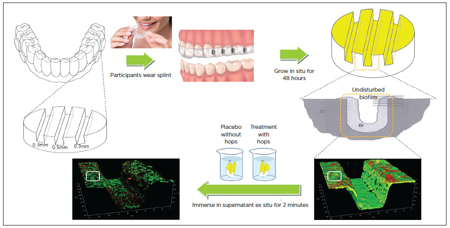
Figure 1

Figure 2
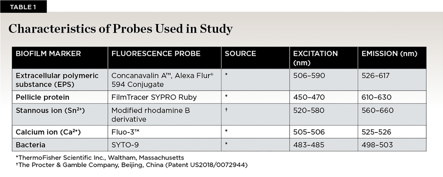
Table 1
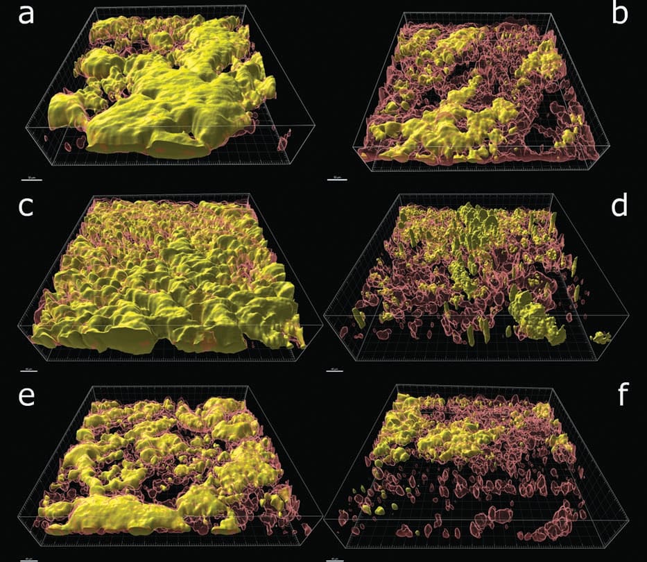
Figure 3
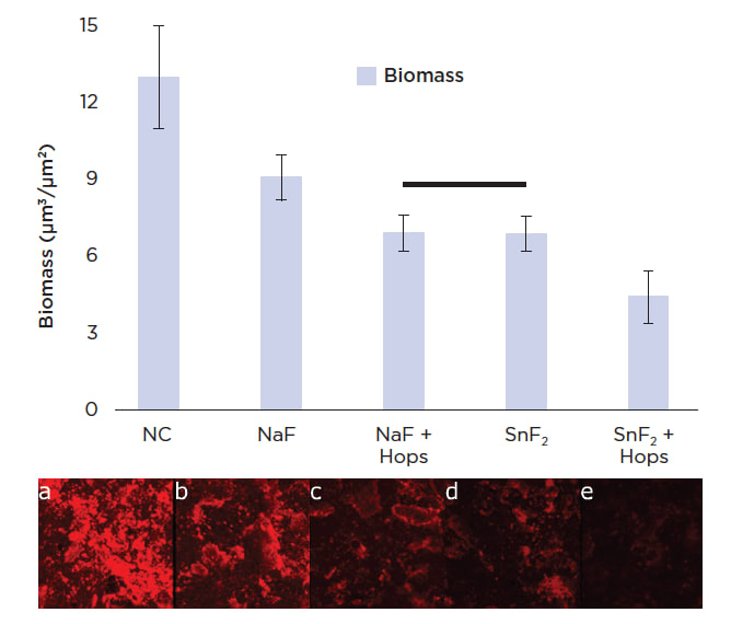
Figure 4
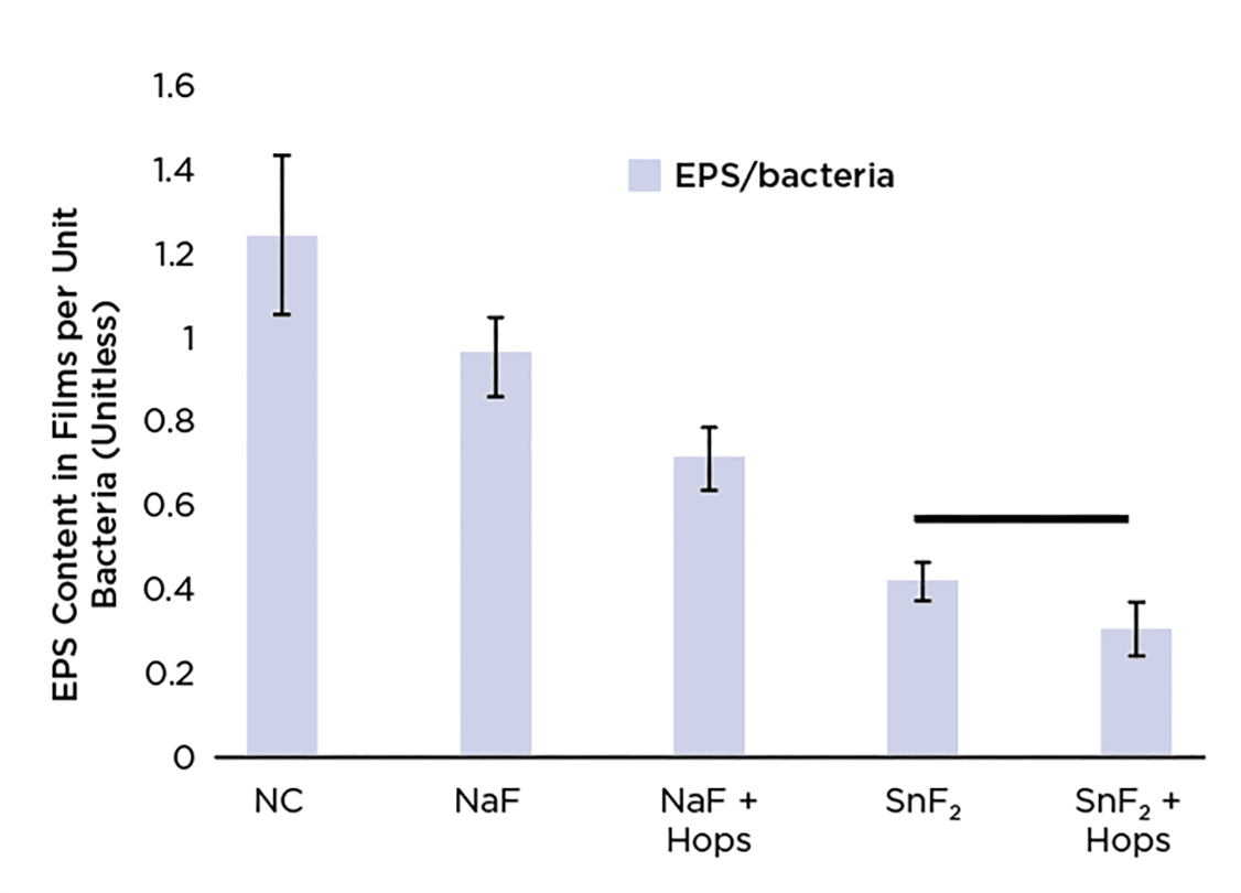
Figure 5

Figure 6
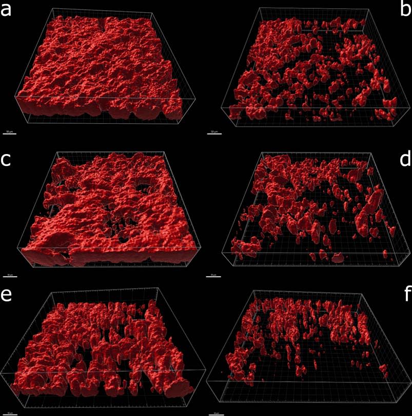
Figure 7
