Cara A. Lund, DMD
A 46-year-old female patient presented with the chief complaint of disliking the shape, color, and spacing of her front teeth. After conducting online research, the patient was adamant she did not want veneers or traditional bonding but instead preferred Bioclear® composite restorations (Bioclear, bioclearmatrix.com) on her maxillary and mandibular anterior teeth to preserve tooth structure. An intraoral scan using the iTero Element® 2 intraoral scanner (Align Technology, Inc, itero.com) and photographs were taken. It was discussed with the patient that prior to final restorations Invisalign® clear aligner therapy (Align Technology, Inc, invisalign.com) was needed to close all mandibular spacing (eliminating the need for restorations) and partially close the maxillary spacing (improving the height-to-width ratios of the final maxillary restorations). The patient completed Invisalign treatment in 15 weeks and then whitened with Opalescence™ PF 10% whitening gel (Ultradent, ultradent.com). An intraoral scan with the iTero Element 2 scanner was taken for a pre-prosthetic lab wax-up of teeth Nos. 6 through 11. These teeth were restored with full esthetic composite veneers using the Bioclear heated composite injection overmolding technique with Filtek™ Supreme Ultra Universal Restorative White Body paste and flowable (3M Oral Care, 3m.com). No tooth structure was resected. The patient was immediately scanned for Vivera® retainers. She was thrilled with her smile transformation.
Key Takeaways
The iTero Element 2 intraoral scanner provides clinicians a formidable system for streamlining workflow, enhancing precision, and connecting more effectively with patients.
Invisalign clear aligner therapy can be utilized to rapidly close spacing and improve height-to-width ratios of final restorations; custom-fit Vivera retainers help maintain teeth alignment.
Direct composite restorations with the Bioclear method
are minimally invasive, additive, and esthetically natural looking.
About the Author
Cara A. Lund, DMD
Private Practice, Stoneham, Massachusetts
Figures and Images

Fig 1

Fig 2

Fig 3
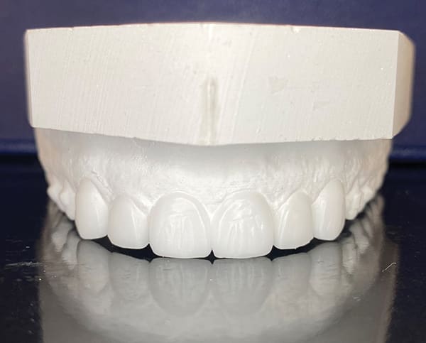
Fig 4
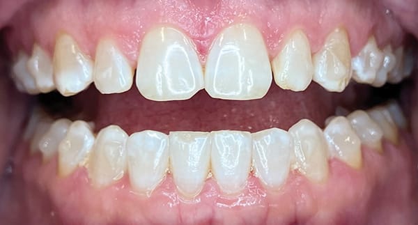
Fig 5
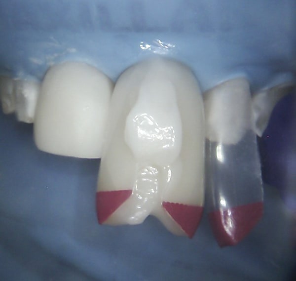
Fig 6
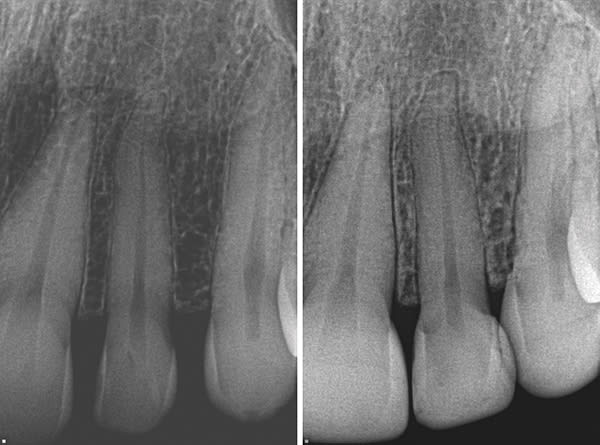
Fig 7
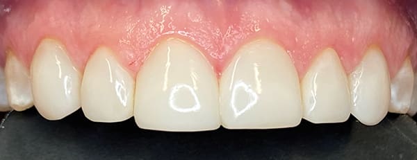
Fig 8

Fig 9
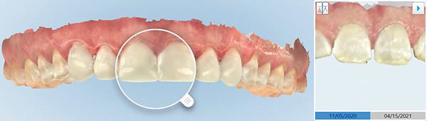
Fig 10
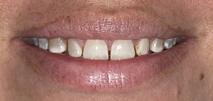
Fig 11
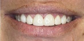
Fig 12