With the application of new products and innovative techniques, extreme clinical cases that in the past required multiple, staged surgical procedures can today be solved with a single-step protocol. In his lecture at the 2018 Southern Implants International Forum, Francesco Amato, DDS, MD, PhD, discussed some of the most anatomically challenging cases and conditions and offered clinical suggestions, practical tips, and scientific evidence for avoiding complications and facilitating predictable results. The main message of his lecture was that practitioners can frequently overcome anatomical limitations through the use of a single, minimally invasive procedure with site-specific implants.
Advanced Sinus Pneumatization
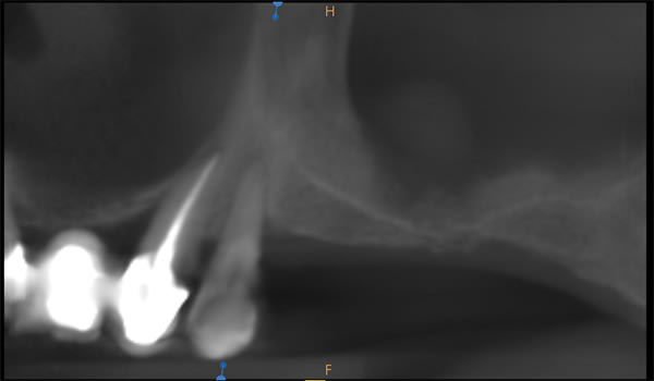
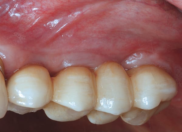
In cases of advanced sinus pneumatization, typically a sinus lift followed by grafting is done to prepare for placement of an implant. This process often requires a longer treatment period and a more invasive procedure than can be achieved with many products and techniques commonly used today. In the case shown (Figure 1 and Figure 2), the patient lacked sufficient bone in the premolar site for placement of an implant of conventional length. By using Southern Implants' Ultra-Short implants, the patient's existing bone could be maximally engaged. Both Ultra-Short implants were 4.5-mm diameter x 4.1-mm long, externally hexed (Figure 3 through Figure 8). Insertion torques of >50 Ncm were achieved.
Ultra-Short implants (≤6 mm) have more than 5 years of follow-up showing a survival rate of 98% in multiple bone classifications, with both immediate and delayed loading in both maxilla and mandible.1
Sinus Floor Proximity
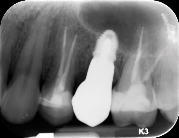
Besides requiring sinus pneumatization, the floor of the sinus can occupy valuable space. In a case with a single posterior tooth extracted and immediately loaded, the tapered Ultra-Short implant was placed (Figure 9 through Figure 15). The domed apical end of the implant prevents penetration of the sinus floor while allowing good primary stability to be achieved. A combination of allograft and xenograft was packed around the implant. Again, insertion torque was >50 Ncm.
Severe Crestal Atrophy
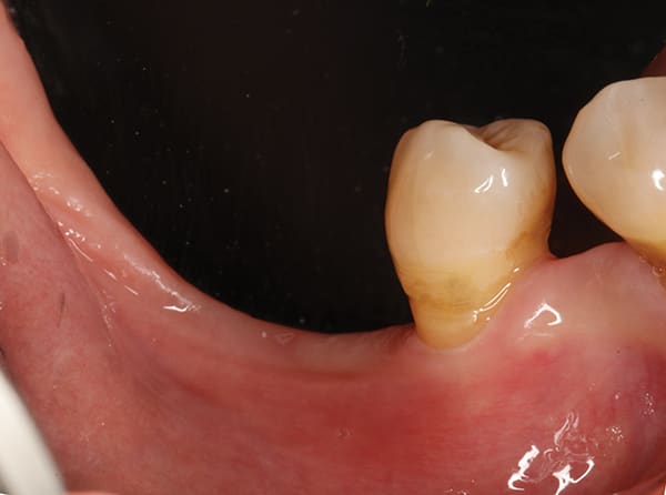
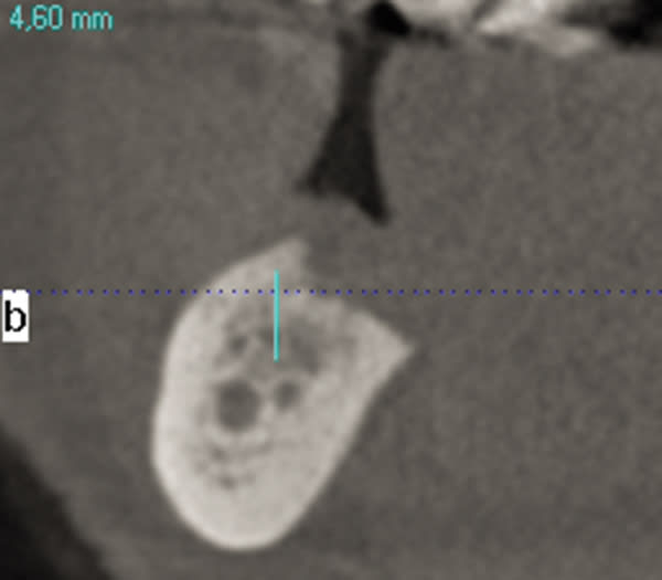
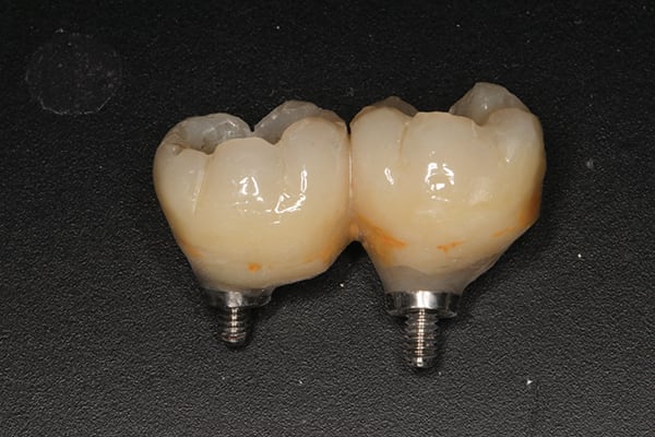
The patient presented with the posterior mandible showing severe atrophy (Figure 16). The computed tomography (CT) scan showed 3.6 mm and 4.6 mm of available bone height with a failing premolar (Figure 17 and Figure 18). To regain function, the premolar was removed, and two Ultra-Short implants were placed in the molar region and splinted together (Figure 19 through Figure 22).
Acute Lesion
Acute lesions are thought to be reasons to use a delayed placement protocol. Use of a dual-axis, subcrestal angle-corrected implant allows the clinician to remove the lesion and atraumatically extract the tooth. This leaves good-quality bone intact while allowing the lesion to heal. The 12° Co-Axis® implant (Southern Implants) is placed into the good bone at an angle while the prosthetic platform still emerges in the correct position, occlusally parallel. As is also the case for standard implants, >70 Ncm insertion torque was readily achieved when all walls of the site were engaged, and >50 Ncm insertion torque was possible in compromised sites with atraumatic extractions using the "ice cream cone" regenerative technique to maintain soft-tissue contours.
Apical Cyst
Molars are the most commonly lost teeth and are frequently subject to apical cysts. With the use of an ultra-wide implant such as the Southern Implants MAX, it is not necessary to delay treatment. After atraumatic removal of the tooth, keeping the interradicular bone intact, the MAX implant is placed immediately with a polyether ether ketone (PEEK) anatomically shaped healing abutment to maintain soft-tissue contours. The MAX implant engages a larger surface area of the socket walls and is placed away from the buccal plate. The sharply tapered body achieves excellent insertion torque. A retrospective study by Amato et al2 has shown survival rates of 99% using wide implants with and without lesions. It is not unusual to reach insertion torques >100 Ncm with ultra-wide implants, as the insertion torque increases with diameter.
Missing Facial Plate
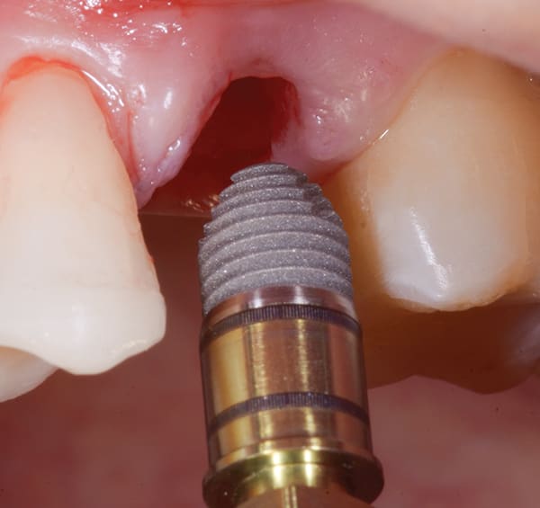
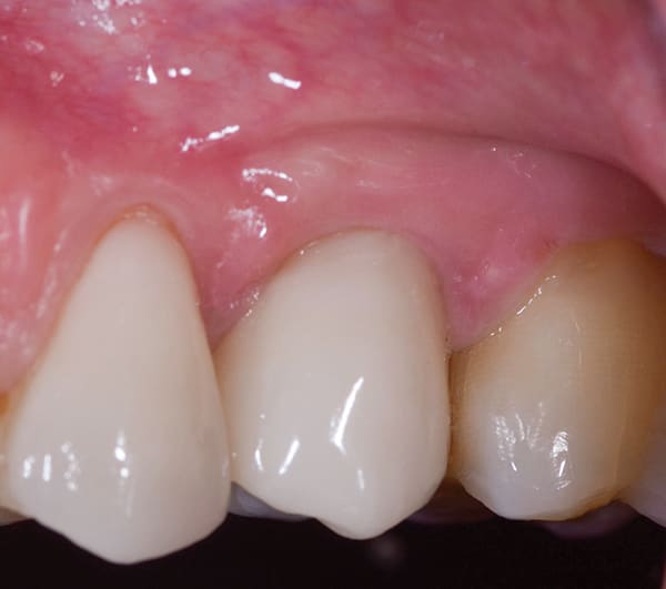
Screw-retained restorations are preferable to cementation for reasons of retrievability and biological complications. Due to the anatomy of the anterior maxilla, angle correction is required for the prosthetic table to be in the correct position to allow screw retention in many cases. Angled abutments increase cost and take up vertical prosthetic space. Using a 24° dual-axis implant (Co-Axis) allows the implant to engage the palatal bone and achieve primary stability regardless of the missing buccal plate. The implant was tightened to 50 Ncm, and the facial plate was grafted with a combination of allograft and xenograft, maintaining tissue contours.
The Presenter
Francesco Amato, DDS, MD, PhD
References
1. Lai HC, Si MS, Zhuang LF, et al. Long-term outcomes of short dental implants supporting single crowns in posterior region: a clinical retrospective study of 5-10 years. Clin Oral Implants Res. 2013;24(2):230-237.
2. Amato F, Polara G. Immediate implant placement in single-tooth molar extraction sockets: a 1- to 6-year retrospective clinical study. Int J Periodontics Restorative Dent. 2018;38(4):495-501.

