Integrating advanced dental technology in everyday dentistry
Dental clinicians hear the term “state-of-the-art” on an almost daily basis. While those in the dental field always aim to offer the latest and greatest, “state-of-the-art” is never static—it is a moving target. Clinical devices, methods, and techniques change so rapidly that it can be a daunting task for practitioners to keep up, let alone become experts regarding what technology defines “state-of-the-art.”
There are many technologies available that enable dentists to deliver the highest level of care for their patients, ranging from diagnostic technologies that visualize soft tissue pathologies to intraoral cameras and diagnostic digital radiographic technologies like digital x-rays. There are also a wide range of computer management-based technologies that not only help clinicians manage logistics within their practices, but also connect to the web for online marketing, data-sharing, and peer-to-peer communication. Some of the latest technologies are treatment-based, like dental lasers, chairside CAD/CAM systems, and advanced dental imaging systems such as cone-beam imaging technology. These innovations empower clinicians to achieve excellence in diagnosis and treatment planning, especially when working with dental implants.
In the author’s estimation, two technologies set the state-of-the-art standard in modern dentistry: CAD/CAM digital impression scanning and cone-beam digital imaging. This article will explore these two technologies, discussing how they enhance the delivery of high-quality patient care by improving clinicians’ ability to diagnose, plan, and treat their patients. What makes these technologies even more exciting is their ability to integrate, allowing dentists to see and do things that were never before imaginable.
Laying the Groundwork
The foundation of restorative dentistry is the restoration of individual teeth. Single-tooth restorative cases are the bread and butter of general restorative dentists. These procedures can include pit and fissure sealants, preventive resin restorations, amalgam restorations, direct and indirect adhesive restorations, cast restorations, CAD/CAM-milled restorations, and stereo-lithographically printed restorations. Dental practitioners have witnessed and participated in the decline of amalgam in restorative dentistry, and have practiced these procedures with all types of materials, such as resin, metal-free ceramic, and hybrids that offer a higher degree of esthetics with superb fit. These materials are all fully capable of meeting patient expectations.
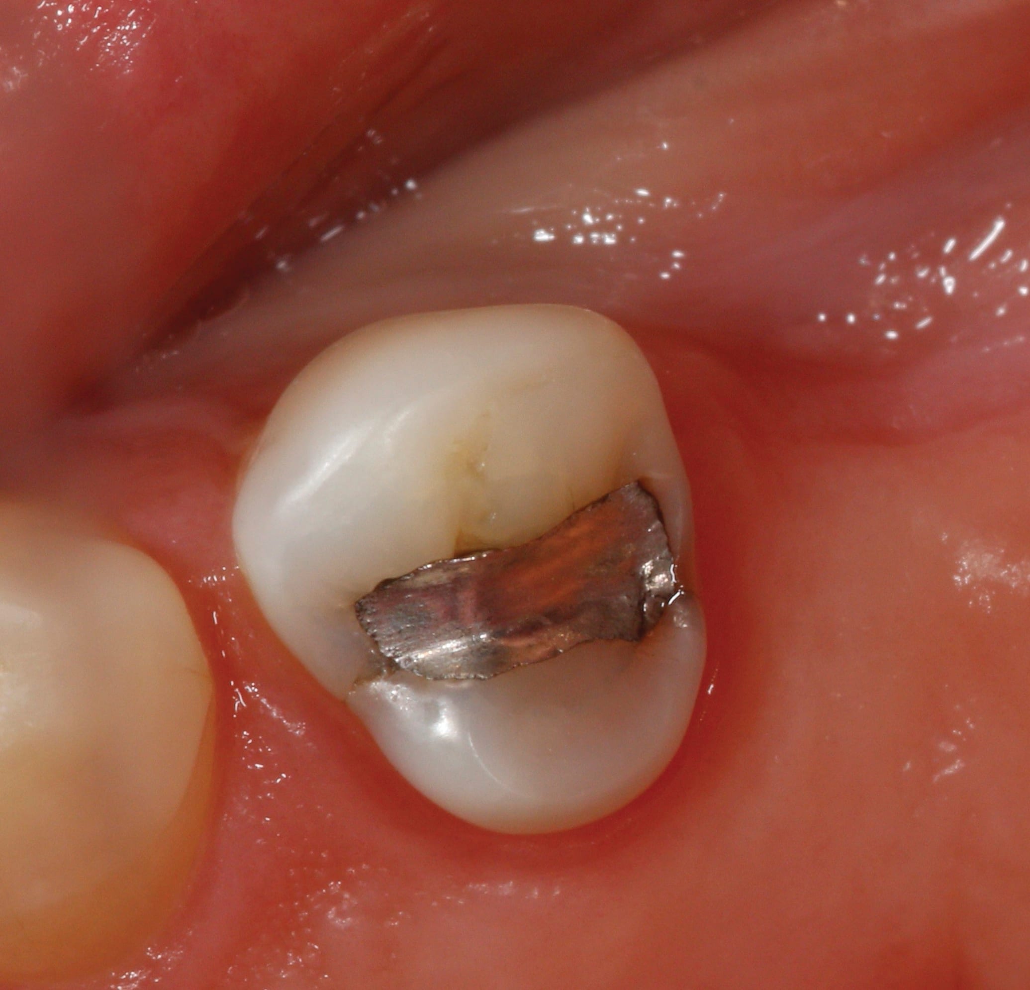
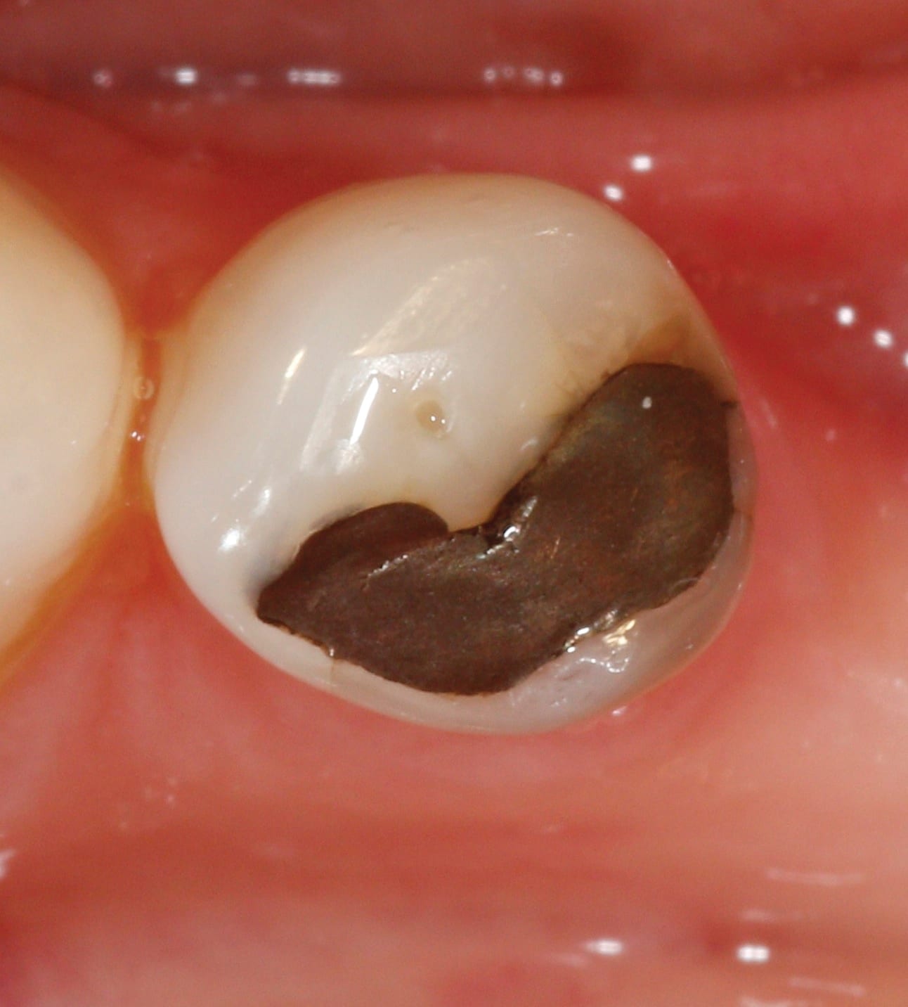
Metal-free restorations are adhesive restorations that allow for a high degree of esthetics and longevity when material selection guidelines are followed and proper operative technique is employed. However, now that amalgam is being edged out of the equation, material choices may become a little more difficult. Planning treatment is simple in situations in which existing amalgam is conservative and tooth structure is sufficient (Figure 1), however, it is much more difficult to plan patient treatment when confronted with a situation in which the existing restoration undermines tooth structure and compromises support (Figure 2).
Dentists need to develop a set of parameters that help them determine which types of materials are best in different clinical situations. There are indications and options for each type of restoration to consider, including the size of the lesion, the size and number of restorations, the location of the restoration margins, the location of the tooth in the arch, and tooth anatomy.
It must be kept in mind that composites take much longer to place than amalgam and that proper placement is highly sensitive to technique. Marginal leakage is a consideration and material wear may also be an issue. Contacts are difficult, especially with larger restorations, and longevity is always a concern, as the durability of composite resins is less than that of amalgam.
When lesions are well contained by tooth structure, composites are the ideal choice. However, when existing defective restorations and carious lesions are too large to be restored directly and full coverage is not warranted, ceramic porcelain inlays and onlays are indicated as the treatment of choice. Important factors to consider when selecting the type of restorative material are the size of the restoration, the opposing dentition, the opposing occlusal pattern, patient habits, patient concerns regarding restorative longevity, and the future likelihood of endodontic involvement (Figure 3). A good rule to follow is that direct composites are not indicated when tooth damage undermines a cusp by extending between half and two-thirds of the way beyond it. In such a case, support should be gained by bonding indirect resin, hybrid, or ceramic materials.
Ideal indirect posterior restorations display several qualities. They offer a good resistance to wear with low abrasion and high hardness. They are also easy to polish and finish, offer a good flexural strength, are reactive to light, and available in varying esthetic shades. When restoring posterior teeth with esthetic materials, clinicians have several choices: direct resin restorations, laboratory-processed ceramic or processed resin restorations, and CAD/CAM chairside fabricated restorations. Excellent results are possible regardless of the method with proper case selection. Laboratory-processed ceramic inlays and onlays, as well as laboratory-processed ceramic crowns, are known to achieve excellent marginal adaptation, well-fitting contacts, well-defined occlusal anatomy, and excellent esthetics (Figure 4).
Despite their potential for an excellent outcome, laboratory-processed restorations also have many drawbacks. They require physical impressions as well as second restorative visit for insertion, which amounts to unproductive chairtime. Patients often feel inconvenienced by this second visit, and the cost of the restoration overall is higher. Provisionals are also required, and creating and fitting them is time-consuming. Additionally, a percentage of these will need to be re-cemented and post-operative discomfort tends to increase in these circumstances. When considering all the facts, the bottom line is that indirect laboratory-processed restorations are time-consuming, inconvenient for patients, and expensive for the practice. In dentistry, time is money.
The Benefits of Digital Impressioning
The most critical step in delivering a well-fitting, indirect restoration is the dental impression. Traditional impression techniques have a long history of success, but also a long list of difficulties. Generally, patient acceptance is poor because these types of impressions commonly cause the patients to gag, may cause tissue abrasion when trays aren’t fitted properly, and most of the materials have an unpleasant taste and consistency. In addition, traditional impression materials are expensive, as are trays, adhesives, impression processing, and infection control procedures. Also, traditional impressions require pouring. If this is delegated to an outside laboratory, impression pouring involves a loss of control over the proper mix ratios and margin marking. When impressions are poured in-office, the office retains control, but staff time is utilized, proper powder-to-water ratios must be observed, and care must be taken to create and mark dies properly. Traditional impression techniques also require a second office visit for restoration seating.
Typically, the required time in the laboratory is two weeks and provisional restorations are needed during that timeframe. Statistically, there are also problems with re-cements and laboratory work not being returned on time. Additionally, there is the cost of laboratory-processed restorations to be considered. There are difficulties related to laboratory prescriptions, along with the time needed for telephone clarification with the laboratory. Remakes involve additional time and costs. Cases also must be catalogued, managed, and stored.
One of the most exciting and promising restorative modalities in dentistry today involves the use of digital impressioning as a replacement for traditional impression materials and trays.
The Planmeca FIT™ system is the leading chairside scanner in the dental marketplace, and it offers the dentist complete control over the entire restorative process. Further benefits include the fact that CAD/CAM restorations conserve tooth structure, and with chairside milling capability, the cases performed with digital impressioning are single-visit procedures that do not require impression materials or trays. Provisionals are not necessary, thus enhancing patient comfort and convenience.
With the the Planmeca FIT™ system, staff participation, interest, and ownership of procedures are increased. The system is fully portable and its architecture is open, meaning that .STL files containing the images can be openly transferred to any milling system. Laboratory costs are also significantly decreased with digital impressioning over conventional impression techniques. The use of digital impression techniques results in a significant reduction of overhead and an increase in profitability when delivering restorative dental care (Figure 5 through Figure 8).
Consider this example. Using a conservative fee of $1,000 per restoration, the profit-per-unit when fabricating restorations chairside with Planmeca FIT™ is $610. If a Planmeca FIT™ system costs $2,400 to lease per month, it would only require creating four-to-five units during that timeframe to cover the cost (Figure 5). With all other factors being equal, when calculating the cost for restorations at $175 per unit for laboratory-made Ivoclar Vivadent IPS e.max® (www.ivoclarvivadent.com) ceramics, the hourly net from fabricating restorations chairside instead of sending them to a laboratory is nearly double when created with Planmeca FIT™ (Figure 6). When taking into account the cost of multiple visits for restorations produced by a laboratory, the profit differential can be conservatively calculated at $32,000 annually (Figure 7). When comparing the average laboratory cost-per-unit and Planmeca FIT™ cost-per-unit for e.max and Ivoclar Vivadent Empress® restorations over the course of a month, the cost of leasing the technology, along with blocks for milling and laboratory fees, are essentially the same (Figure 8).
Digital impressions with Planmeca FIT™ are proven to fill a void in the current system of planning and delivering dental treatments. They allow for conservative treatment and the marginal integrity of restorations that are equal to or better than what can be accomplished by commercial laboratories. The Planmeca FIT™ is designed around the simple portability of a laptop and a wireless connection. Its scanner uses blue laser pattern triangulation to capture an image accurately and efficiently. The scanner tips are also autoclavable. Once images are acquired, they are interpreted and displayed using the powerful and intuitive Planmeca Romexis® software. With Planmeca Romexis software, there is full integration with Planmeca 3D-imaging. In fact, Planmeca Romexis is designed with open architecture that allows the import and integration of all .STL and .DCM images from any system in the marketplace and does not restrict its imaging to its own propriety software. This system utilizes the powerful, fast, and advanced milling of a wide range of materials with the PlanMill™ 40 milling system.
Planmeca FIT’s™ chairside milling materials include Ivoclar Vivadent’s e.max® CAD LT and HT and IPS Empress, Lava Ultimate™ by 3M ESPE (www.3mespe.com), Ivoclar TelioCAD provisonal material, and 3M Paradigm MZ100 blocks.
The benefits of digital impressioning are numerous. First, there is no need for trays, adhesives, or impression materials. Models do not need to be poured, and when retakes are necessary they can be achieved instantaneously. Margins are marked immediately. The time needed to create an accurate model and articulation is significantly reduced while patient comfort is significantly enhanced. Infection control protocols are streamlined. Perhaps most importantly, accuracy is inherent in digital impressioning, and complete control is directly in the dentist’s hands.
Digital impressioning represents a new dawn in dentistry. It alters the traditional workflow and directly yields greater consistency of quality results while enhancing patient comfort and convenience. It also generates time-savings, significant financial savings, and decreased time for restoration. In fact, digital imaging allows clinicians to create ceramic, hybrid ceramic, resin, and provisional restorations chairside within appointment times of as little as 60 minutes.
This case (Figure 9 through Figure 12) illustrates the benefits of digital dentistry in terms of saving time with an esthetic ceramic restoration fabricated in a single visit, patient convenience, quality of the restorative result, and a high return on investment.
About the Author
Eugene Antenucci, DDS, FAGD, DICOI, serves as Assistant Clinical Professor at NYU College of Dentistry, and maintains a full-time private practice in Huntington, NY.


