ABSTRACT
In the posterior maxilla, bone volume, height, and density are often insufficient for implant placement and rehabilitation, which can affect implant long-term stability and success. These anatomical limitations may dictate the need for sinus grafting procedures. Osseodensification is an implant site instrumentation method that enhances bone density and can also be used for transcrestal maxillary sinus augmentation. This technique utilizes bone plasticity through specially designed burs that compact and densify bone along the osteotomy walls while simultaneously propelling bone particles laterally and apically. This process generates a hydrodynamic compaction wave at the bur tip, which propels irrigation fluid into the sinus cavity lifting the membrane and simultaneously compacting bone particles grafting the sinus with autogenous bone. Additionally, the flutes of the osseodensification bur permit the operator to subsequently graft biomaterial, facilitating further elevation of the Schneiderian membrane. Current literature outlines three specific protocols for sinus floor elevation and implant placement utilizing osseodensification with residual bone height (RBH) requirements: ≥6 mm for protocol I, ≥4–5 mm for protocol II, or ≥2–3 mm for protocol III, with concurrent implant placement in the presence of adequate bone and soft-tissue volume. Osseodensification-mediated sinus grafting allows for shorter surgery duration, reduced postoperative edema, less reported pain, and subsequently decreased analgesic intake compared to lateral window techniques. This article details the step-by-step osseodensification sinus lift protocol IV, a two-stage sinus floor elevation indicated in cases of RBH ≤0.5–1.5 mm as an alternative to lateral window techniques.
Inadequate bone volume and density are frequent challenges observed in the implant rehabilitation of partially or fully edentulous patients. Alveolar bone atrophy and pneumatization of the maxillary sinus followed by tooth extraction in the maxilla may limit the possibility for the placement of implants or for adequate bone regeneration combined with implant placement.1 Maxillary sinus floor augmentation via a lateral window, Schneiderian membrane elevation, and creation of space for bone augmentation is well documented.2,3 This technique, however, requires a wide mucoperiosteal flap, significant lateral opening preparation, and membrane separation, making the procedure complex with a reportedly high membrane perforation rate of up to 44% in the conventional approach and increased to 56.4% when lateral wall thickness was ≥2 mm.4-6 In addition to bleeding, these intraoperative complications are the most common problems postoperatively and may lead to sinus graft infections, delayed healing, and postoperative discomfort.7 Patients with severe maxillary bone deficiency, unfavorable sinus septum anatomy, and thick lateral buccal bone present a significant challenge.
Alternative access, therefore, can be created by the direct palatal approach or crestal approach.8-10 The palatal approach, however, is also associated with several significant clinical limitations, including restricted access, limited visual field, the need for specially engineered curved instruments, and the anatomical proximity to the greater palatine artery. Additionally, a thermoformable appliance or full denture used by edentulous patients to provide palate flap stabilization are also required.11
The less invasive crestal approach was introduced by Summers who used osteotomes for both sinus floor elevation and ridge expansion. Literature reported that patient satisfaction was higher due to the shorter operation time and smaller wound.12 Historically, the crestal approach method was recommended in cases with >5 mm of residual bone height (RBH) and a flat sinus floor,13,14 and because of implant failure rate, it was linked directly to initial RBH of <4 mm.15 Other reports indicated higher success rates with two sequential combined lift with osteotome and balloon approaches were used in cases of RBH of >3 mm.16 However, tapping bone with different densities with osteotomes may create uncontrolled fracture of the sinus floor or provoke benign paroxysmal positional vertigo.17 The piezoelectric technique with special sinus lift tips was reported to effectively lift the membrane with the hydraulic pressure of a physiologic solution subjected to piezoelectric cavitation; however, this method still requires a lateral window with significant additional chairtime and initial removal of the patient’s own vital bone to create the access to the Schneiderian membrane.18
It can be concluded that both the crestal and lateral approaches have high success rates; however, crestal techniques are usually shorter in time and less invasive.19,20 Dental implants placed using both techniques in grafted sinuses are considered effective and safe for rehabilitation, with success rates surpassing 93% for lateral window and 97% for transalveolar approaches in medium- and long-term observations.21 There were no statistically significant differences found between implants placed using one-stage versus two-stage sinus lift techniques. Several studies, however, indicated that in patients with a residual bone height of 1 mm to 3 mm beneath the maxillary sinus, the one-stage lateral sinus lift approach may carry a slightly increased risk of implant failure.22 Therefore, implants should not be placed in one-stage approaches simultaneously with the sinus lift procedure when RBH is <1–2 mm.
Novel Bone Instrumentation Technique
A novel bone instrumentation technique was introduced utilizing specially designed tapered fluted burs.23 Osseodensification burs (Densah® burs, Versah LLC, versah.com) used for implant site instrumentation produce bone compaction–autografting and preserve bone bulk, accelerating bone remodeling and regeneration.23-25 Furthermore, in the posterior maxilla, this technique progressively moves autogenous bone apically into the sinus cavity and into the trabecular space. Osseodensification burs can be used in clockwise (CW) and counterclockwise (CCW) directions and provide less vibration than regular implant drills.23,26,27
The ability of the osseodensification process to elevate the sinus floor without perforating the membrane relies on the CCW action of the densifying burs. This motion enhances irrigation throughout the osteotomy, maintaining fluid presence at the instrument tip. As a result, once the sinus floor is engaged, the irrigation and autogenous bone chips create a hydraulic wave that gently separates and lifts the sinus membrane off the bony bed and, simultaneously, autografting bone.28 Osseodensification has been documented with long-term data as safe and predictable for sinus elevation in cases with limited residual bone height.28-30 The reported minimum bone height required remains unclear in the current literature.30,31 Multicenter clinical studies confirmed the predictable outcome of osseodensification for transcrestal sinus floor elevation, even in cases with low crestal bone height, with a low rate of membrane perforation. Severe posterior maxillary bone loss, however, was identified as a risk factor for higher perforation rates.32 While these findings offer important data for clinical practice, further evidence is needed to evaluate the long-term outcomes of implants placed in sinus-augmented sites with <2 mm crestal bone height using this transcrestal osseodensification protocol.
This article aims to present the efficacy of osseodensification instrumentation for cases with severe crestal bone loss of 0.5 mm to 1.5 mm as a two-stage approach for implant placement and rehabilitation of the posterior maxilla.
Osseodensification Sinus Graft Protocol IV
Ridge width must be >7 mm. It is recommended to limit the introduction of the initial Densah bur up to 1 mm beyond the sinus floor. The burs must never advance more than 1 mm to 2 mm beyond the sinus floor, regardless of bur diameter or stage (Figure 1). Use Versah Vertical Stops to control entry depth (Figure 2, right).
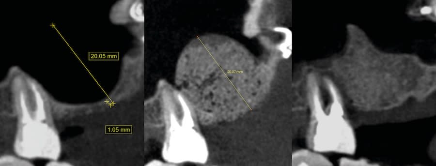
Step 1: Measure bone height at the osteotomy site on the CBCT. Measure ridge clinical width. A minimum of 7 mm alveolar ridge width is needed. Perform horizontal incision 2 mm to 3 mm palatally from the planned osteotomy site and elevate the flap using regular techniques of the clinician’s choice.
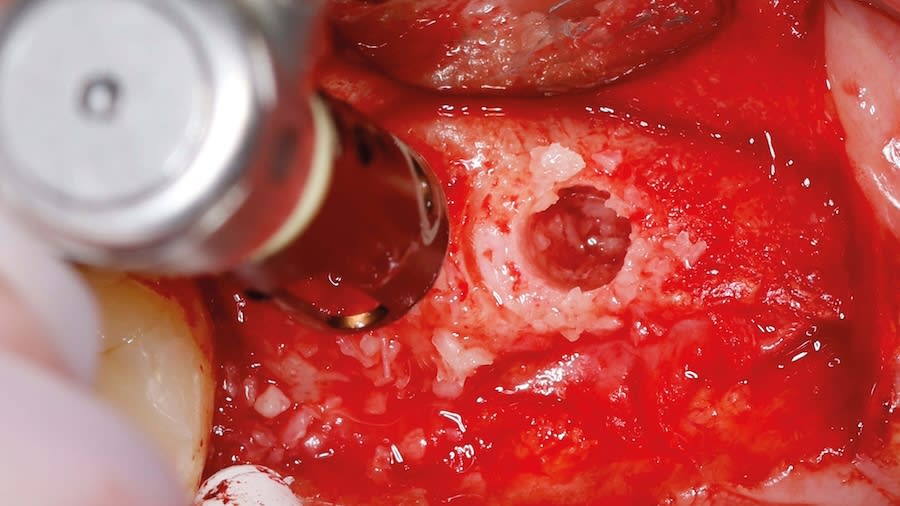
Step 2: Use Densah bur VT4555 (Figure 2, left) in CCW mode in 1 mm increments up to 2 mm past the sinus floor. Use Vertical Stops(Figure 2, right) to control entry depth.
Avoid using pilot and small-diameter burs. Start with Densah bur VT4555 (5.0) in CCW mode with Vertical Stop to reach the sinus floor.
Use surgical motor in reverse-counterclockwise at 1,000 RPM to 1,100 RPM with constant irrigation in all osteotomies. Keep running the bur in the osteotomy until reaching the sinus floor, then the membrane, and subsequently pass 1 mm increments up to a maximum 2 mm past the sinus floor with a modulating pressure in a pumping motion (cortical sinus floor level is determined on initial CBCT measurements). Densah bur VT4555 (5.0) could be used with a modified dull tip. Use Vertical Stop (Figure 2, right) to facilitate the need for safe controlled advancement (Versah Sinus Kit, Versah LLC).
Step 3: Graft placement: Once the final osteotomy/lift is reached, it is suggested to place one platelet-rich fibrin (PRF) membrane (cut into three smaller pieces) or two wet soft hemostatic sponges to be pushed gently with the Densah bur (5.0) before introducing well-hydrated particulate allograft or biomaterials.
Use same Densah bur VT4555 (5.0) with Vertical Stop in the CCW motion at low speed (100 RPM) without irrigation and gently advance it 1 mm to 2 mm to propel the allograft into the sinus. Repeat the graft compaction step to achieve further elevation of the sinus membrane. Keep propelling well-hydrated allograft or other biomaterial until achieving adequate grafting volume. Achieve full adequate flap primary closure.
Step 4: Allow 4 to 6 months for the sinus graft healing. CBCT evaluation is needed to evaluate the available regenerated bone height for the implant placement. Reflect soft tissue utilizing standard surgical instruments, and use Densah burs for osseodensification site preparation to prepare the implant osteotomy and place the implant.
In cases where additional bone lift is required in stage two, apply osseodensification sinus grafting protocols (Versah) I or II as needed prior to implant placement.
Three cases demonstrating the osseodensification sinus graft protocol 1V are presented.
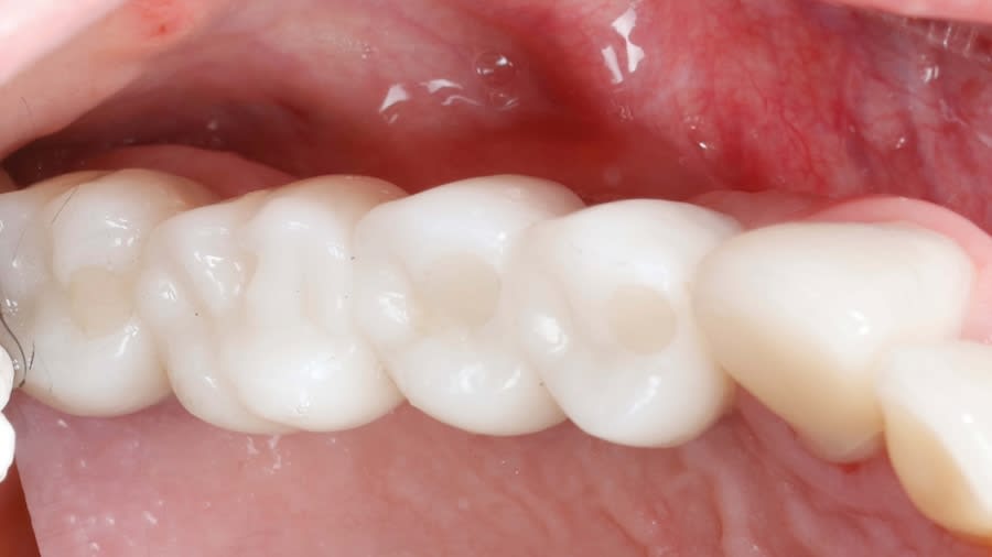
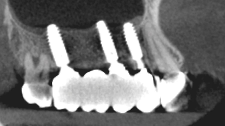
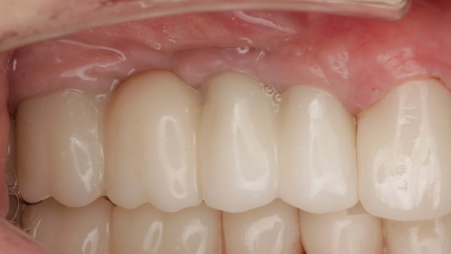
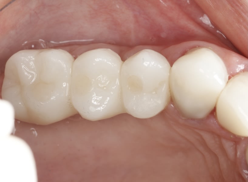
Case 1 (Figure 3 through Figure 24) depicts a 3-year follow-up of combined osseodensification sinus protocol IV in a severely resorbed maxillary ridge with ≤0.5 mm bone height in molar sites and horizontal deficiency at the first premolar site, using a two-stage approach for implant placement.
Case 2 (Figure 25 through Figure 36) illustrates a 3-year follow-up of the osseodensification sinus protocol IV in a severely resorbed right maxillary ridge with <0.5 mm bone height in molar sites, using a two-stage approach for implant placement.
Case 3 (Figure 37 through 44) shows a case of significant trauma history with a 3-year follow-up of the osseodensification sinus protocol IV in a severely resorbed right maxillary ridge with ≤0.5 mm bone height in molar sites, using a two-stage approach for implant placement.
Discussion
Sinus floor elevation and bone augmentation is a common procedure used to augment bone height in the posterior maxilla for implant-based prosthetic reconstruction. The earlier and more teeth are lost the more bone bulk loss is observed.33
Using short implants in the atrophic posterior low bone quality maxilla is not recommended.34,35 Historically, posterior maxilla sinus vertical augmentation through the use of osteotome or other crestal methods has significant limitation directly related to the initial autogenous bone volume that can be pushed and moved toward the sinus without additional biomaterial.12,23 Pterygoid and zygomatic implant treatment modalities have been introduced to avoid sinus grafting but have their own challenges.36-39 Implant placement with sinus augmentation shows reliable long-term survival outcomes and is still considered as the gold standard.40
Sinus floor elevation techniques and instrumentation improvements allowed practitioners to choose the proper surgical approach according to the anatomical factors of the case.8,41-43 Studies suggest that when ridge height is severely reduced <2 mm, a lateral window approach is indicated.19 The meta-analysis of published data between 1980-2016 shows that the high perforation rate in lateral sinus floor augmentation can be reduced by using piezoelectric tips instead of high-speed rotary instruments; however, the lateral window method in these cases still requires large flap elevation and longer surgery time.44 Additional factors such as smoking, thin Schneiderian membrane, irregular or narrow-tapered sinus anatomy, and the presence of sinus septa are associated with increased risk of membrane perforation during the lateral elevation procedures,45 with membrane perforation leading to graft migration or loss, infection, and procedure failure.46,47
Understanding the structural properties of the Schneiderian membrane to achieve tension lower than 7.3N/mm³ and stretching less than 132.6% (in one dimension) and 124.7% (in two dimensions) could also potentially lower its perforation complication frequencies.48 Normal membrane thickness in healthy patients is considered to be 1 mm, however 3-dimensional imaging techniques tend to overestimate its thickness by 2.5 times compared to histologic evaluation.49 Perforation rate in transcrestal sinus approaches has been reported to be higher when the membrane is thicker than 3 mm or thinner than 0.5 mm. Periodontitis and smoking may result in thickening of the sinus membrane and cause it to be more prone to perforation. It was suggested that people >40 years old tend to have thicker membranes (2.17 mm ± 1.9 mm) when compared to patients <40 years old (1.32 mm ± 1.09 mm), but no correlation was found between sex or season.50
Osseodensification used for the transcrestal sinus floor elevation resulted in a 97% implant survival rate in cases with crestal bone height between 3 mm and 7 mm.28 This technique takes advantage of specially designed burs (ie, Densah burs) that create hydraulics within the osteotomy. This generates a hydrodynamic compression wave ahead of the bur tip, driving irrigation fluid into the site and compacting autogenous bone particles apically.23,51 The autogenous bone with fluid mixtures generates gentle pressure against the Schneiderian membrane, lifting it and simultaneously placing the bone graft material between the membrane and sinus floor.32 The osseodensification sinus protocol enables the achievement of four objectives using a single instrument, allowing for simultaneous preparation of the osteotomy, reaching the sinus membrane, lifting the membrane off the bony bed, and depositing bone graft material.
A prospective comparative clinical study found that the lateral window technique resulted in four times higher membrane perforation rate than the osseodensification transcrestal approach.31 Another multicenter clinical study reported that in 670 osseodensification sinus grafting procedures in 621 patients receiving implants in cases with remaining alveolar bone ranging from 2 mm to 7 mm, overall sinus membrane perforation rate was 7.31%.32
The selection of bone grafting material for the sinus augmentation has long been a subject of debate, with differing opinions frequently expressed by researchers. Systematic reviews find that allograft bone is effective in sinus floor augmentation, but direct comparisons to xenograft or alloplast are limited.52 Histological and histomorphometry analyses demonstrated that a combination of cortical and cancellous mineralized bone allograft effectively facilitated de novo bone formation and may serve as a reliable grafting material for maxillary sinus augmentation procedures.53 In a recent randomized clinical study on alveolar ridge preservation employing various grafting materials, the allograft yielded the highest proportion of vital bone and the lowest quantity of residual graft particles. Both the allograft and alloplastic materials resulted in significantly greater new bone formation compared to the xenograft. In contrast, the xenograft presented the highest level of residual particles, a value markedly exceeding that observed in the other groups.54 Systematic reviews show that xenografts and alloplasts produce high levels of residual particles of over 35%, and allografts between 12% and 21%, at 7 months. More vital bone was also present at 3 months with allograft, while xenografts showed lower percentage of vital bone at 4 to 5 months.55,56 Additional available evidence indicates that xenograft may remain detectable within the regenerated tissue for as long as 20 years postoperatively.57 In a split‐mouth trial combining graft with autogenous bone, allograft composites remodeled faster than bovine xenograft composites: allograft mix had significantly fewer residual particles and faster turnover than the delayed remodeling in the xenograft group.58
Scientific evidence has concluded that allograft seems to enhance vital bone formation faster than other biomaterials and provide high implant success, especially when combined with autogenous bone compaction autografting provided by osseodensification techniques.23,24 The evidence suggests that the most significant resorption of an allograft occurs within the first 6 months after grafting, with a slower rate of change thereafter.59 A study examining a 1:1 mixture of cortical and cancellous mineralized bone allograft revealed significant results for sinus augmentation procedures. Histomorphometry evaluation at 6 months showed an average vital bone content of 30.32% (25%–44%), with residual non-vital bone of only 18.06%.53 A retrospective study of 39 patients undergoing crestal window sinus elevation with mineralized human bone allografts found that narrower and shorter maxillary sinuses yielded significantly greater high-quality bone formation. Larger sinus dimensions were associated with reduced bone gain highlighting the importance of 3D sinus morphology in planning sinus augmentation procedures.60
Alloplastic putty bone can also be used as a grafting material with the ability to form new bone (47.15% ± 8.5% at 4 to 6 months).61 However, due to its long-term remodeling time and its putty form with low compaction abilities, it might produce several clinical challenges in severe maxillary bone atrophy sites cases with this osseodensification sinus graft protocol IV.
Other studies confirm that wide sinuses in the buccopalatal distances have smaller angiogenesis and lower migration of cells involved in osteogenesis, resulting in lower vital bone formation.62 Therefore, as indicated in the present study, for the two-stage approaches for severely resorbed and wide sinuses, the 4- to 6-month mark appears to be a suitable timepoint for second-stage implant placement post grafting, as this allows for notable graft remodeling while minimizing additional resorption that might occur with longer waiting periods.
Conclusion
The presented protocol describes a crestal sinus elevation procedure using osseodensification burswith vertical stops (Versah) in severe cases with <2 mm of residual bone as a predictable technique, with successful bone regeneration for second-stage implant placement with subsequent stable peri-implant bone level at 3-year follow-up. This technique, thus, offers a simplified, minimally invasive, and less traumatic method for sinus floor elevation in cases with severe residual bone height loss.
DISCLOSURE
The authors had no disclosures or conflicts of interest to report. Dr. Huwais is the developer of the Osseodensification method and inventor of the Densah bur technology used in the case reports.
ABOUT THE AUTHORS
Marcel Firlej, DDS, PhD
Teaching Assistant, Department of Orthodontics and Craniofacial Anomalies, Poznan University of Medical Sciences, Collegium Maius, Poznan, Poland
Salah Huwais, DDS
Clinical Assistant Professor, Department of Periodontics, College of Dentistry, University of Illinois at Chicago, Chicago, Illinois
Rodrigo Neiva, DDS, MS
Adjunct Clinical Professor, Department of Periodontology and Oral Implantology, Kornberg School of Dentistry, Temple University, Philadelphia, Pennsylvania
References
1. Mohan N, Wolf J, Dym H. Maxillary sinus augmentation. Dent Clin North Am. 2015;59(2):375-388.
2. Tatum H Jr. Maxillary and sinus implant reconstructions. Dent Clin North Am. 1986;30(2):207-229.
3. Boyne PJ, James RA. Grafting of the maxillary sinus floor with autogenous marrow and bone. J Oral Surg. 1980;38(8):613-616.
4. Lyu M, Xu D, Zhang X, Yuan Q. Maxillary sinus floor augmentation: a review of current evidence on anatomical factors and a decision tree. Int J Oral Sci. 2023;15(1):41.
5. Basma H, Saleh I, Abou-Arraj R, et al. Association between lateral wall thickness and sinus membrane perforation during lateral sinus elevation: a retrospective study. Int J Oral Implantol (Berl). 2021;14(1):77-85.
6. Schwartz‐Arad D, Herzberg R, Dolev E. The prevalence of surgical complications of the sinus graft procedure and their impact on implant survival. J Periodontol. 2004;75(4):511-516.
7. Testori T, Weinstein T, Taschieri S, Wallace SS. Risk factors in lateral window sinus elevation surgery. Periodontol 2000. 2019;81(1):91-123.
8. Jadach R, Asa’ad F, Rasperini G, Osypko K. Classifying maxillary sinuses of Polish patients for sinus lift: a pilot study. Dent J (Basel). 2024;12(2):35.
9. Jensen OT, Perkins S, Van de Water FW. Nasal fossa and maxillary sinus grafting of implants from a palatal approach: report of a case. J Oral Maxillofac Surg. 1992;50(4):415-418.
10. Rahpeyma A, Khajehahmadi S. Indications for palatal sinus lift: case series. J Indian Soc Periodontol. 2018;22(3):254-256.
11. Stübinger S, Saldamli B, Seitz O, et al. Palatal versus vestibular piezoelectric window osteotomy for maxillary sinus elevation: a comparative clinical study of two surgical techniques. Oral Surg Oral Med Oral Pathol Oral Radiol Endod. 2009;107(5):648-655.
12. Summers RB. A new concept in maxillary implant surgery: the osteotome technique. Compendium. 1994;15(2):152-158.
13. Zitzmann NU, Schärer P. Sinus elevation procedures in the resorbed posterior maxilla. Comparison of the crestal and lateral approaches. Oral Surg Oral Med Oral Pathol Oral Radiol Endod. 1998;85(1):8-17.
14. Rosen PS, Summers R, Mellado JR, et al. The bone-added osteotome sinus floor elevation technique: multicenter retrospective report of consecutively treated patients. Int J Oral Maxillofac Implants. 1999;14(6):853-858.
15. Călin C, Petre A, Drafta S. Osteotome-mediated sinus floor elevation: a systematic review and meta-analysis. Int J Oral Maxillofac Implants. 2014;29(3):558-576.
16. Kfir E, Kfir V, Mijiritsky E, et al. Minimally invasive antral membrane balloon elevation followed by maxillary bone augmentation and implant fixation. J Oral Implantol. 2006;32(1):26-33.
17. Peñarrocha-Diago M, Rambla-Ferrer J, Perez V, Pérez-Garrigues H. Benign paroxysmal vertigo secondary to placement of maxillary implants using the alveolar expansion technique with osteotomes: a study of 4 cases. Int J Oral Maxillofac Implants. 2008;23(1):129-132.
18. Vercellotti T, De Paoli S, Nevins M. The piezoelectric bony window osteotomy and sinus membrane elevation: introduction of a new technique for simplification of the sinus augmentation procedure. Int J Periodontics Restorative Dent. 2001;21(6):561-567.
19. Lyu M, Xu D, Zhang X, Yuan Q. Maxillary sinus floor augmentation: a review of current evidence on anatomical factors and a decision tree. Int J Oral Sci. 2023;15(1):41.
20. Al-Almaie S, Kavarodi AM, Alorf A, Alzahrani S. A split-mouth design comparison for lateral and crestal sinus lift techniques with dental implants placements: short communication. Open Dent J. 2017;11:603-608.
21. Del Fabbro M, Wallace SS, Testori T. Long-term implant survival in the grafted maxillary sinus: a systematic review. Int J Periodontics Restorative Dent. 2013;33(6):773-783.
22. Felice P, Pistilli R, Piattelli M, et al. 1-stage versus 2-stage lateral sinus lift procedures: 1-year post-loading results of a multicentre randomised controlled trial. Eur J Oral Implantol. 2014;7(1):65-75.
23. Huwais S, Meyer EG. A novel osseous densification approach in implant osteotomy preparation to increase biomechanical primary stability, bone mineral density, and bone-to-implant contact. Int J Oral Maxillofac Implants. 2017;32(1):27-36.
24. Lahens B, Neiva R, Tovar N, et al. Biomechanical and histologic basis of osseodensification drilling for endosteal implant placement in low density bone. An experimental study in sheep. J Mech Behav Biomed Mater. 2016;63:56-65.
25. De Carvalho Formiga M, Grzech-Leśniak K, Moraschini V, et al. Effects of osseodensification on immediate implant placement: retrospective analysis of 211 implants. Materials (Basel). 2022;15(10):3539.
26. Slete FB, Olin P, Prasad H. Histomorphometric comparison of 3 osteotomy techniques. Implant Dent. 2018;27(4):424-428.
27. Firlej M, Schwimer C, Martin R, et al. Guided selective preservation of tooth (SPOT) protocol: fully guided socket-shield technique. Compend Contin Educ Dent. 2025;46(2):84-93.
28. Huwais S, Mazor Z, Ioannou AL, et al. A multicenter retrospective clinical study with up-to-5-year follow-up utilizing a method that enhances bone density and allows for transcrestal sinus augmentation through compaction grafting. Int J Oral Maxillofac Implants. 2018;33(6):1305-1311.
29. Orth CC, da Silva RC, de Barros Carrilho GP, et al. Vertical bone gain post‐sinus lifting and simultaneous implant placement with osseodensification: a retrospective study. Clin Implant Dent Rel Res. 2025;27(2):e13430.
30. Gaspar J, Mazor Z, Bonfante EA. Osseodensification technique in crestal maxillary sinus elevation – a narrative review. Clin Implant Dent Relat Res. 2025;27(1):e13399.
31. Gaspar J, Botelho J, Proença L, et al. Osseodensification versus lateral window technique for sinus floor elevation with simultaneous implant placement: a randomized clinical trial on patient‐reported outcome measures. Clin Implant Dent Relat Res. 2024;26(1):113-126.
32. Mazor Z, Gaspar J, Silva R, et al. Maxillary sinus membrane perforation rate utilizing osseodensification-mediated transcrestal sinus floor elevation: a multicenter clinical study. Clin Implant Dent Relat Res. 2024;26(6):1172-1180.
33. Al-Askar M, O’Neill R, Stark PC, et al. Effect of single and contiguous teeth extractions on alveolar bone remodeling: a study in dogs. Clin Implant Dent Relat Res. 2013;15(4):569-575.
34. Jaffin RA, Berman CL. The excessive loss of Branemark fixtures in type IV bone: a 5-year analysis. J Periodontol. 1991;62(1):2-4.
35. Friberg B, Jemt T, Lekholm U. Early failures in 4,641 consecutively placed Brånemark dental implants: a study from stage 1 surgery to the connection of completed prostheses. Int J Oral Maxillofac Implants. 1991;6(2):142-146.
36. Raouf K, Chrcanovic BR. Clinical outcomes of pterygoid and maxillary tuberosity implants: a systematic review. J Clin Med. 2024;13(15):4544.
37. George P, Kurtzman GM. Pterygoid implants: anatomical considerations and surgical placement. J Osseointegration. 2022;14(2):81-87.
38. Tzerbos F, Bountaniotis F, Theologie-Lygidakis N, et al. Complications of zygomatic implants: our clinical experience with 4 cases. Acta Stomatol Croat. 2016;50(3):251-257.
39. Aparicio C, Olivo A, De Paz V, et al. The zygoma anatomy-guided approach (ZAGA) for rehabilitation of the atrophic maxilla. Clin Dent Rev [Internet]. 2022;6(2). https://link.springer.com/10.1007/s41894-022-00116-7. Accessed November 12, 2025.
40. Thoma DS, Cha JK, Jung UW. Treatment concepts for the posterior maxilla and mandible: short implants versus long implants in augmented bone. J Periodontal Implant Sci. 2017;47(1):2-12.
41. Liu Y, Ji P, Fu G, Huang H. Transcrestal sinus augmentation with simultaneous implant placement in 1 to 2 mm residual alveolar bone: a case report. J Oral Implantol. 2022;48(4):319-323.
42. Pignaton TB, Wenzel A, de Almeida Ferreira CE, et al. Influence of residual bone height and sinus width on the outcome of maxillary sinus bone augmentation using anorganic bovine bone. Clin Oral Implants Res. 2019;30(4):315-323.
43. Zhou W, Wang F, Magic M, et al. The effect of anatomy on osteogenesis after maxillary sinus floor augmentation: a radiographic and histological analysis. Clin Oral Investig. 2021;25(9):5197-5204.
44. Jordi C, Mukaddam K, Lambrecht JT, Kühl S. Membrane perforation rate in lateral maxillary sinus floor augmentation using conventional rotating instruments and piezoelectric device—a meta-analysis. Int J Implant Dent. 2018;4(1):3.
45. Shao Q, Li J, Pu R, et al. Risk factors for sinus membrane perforation during lateral window maxillary sinus floor elevation surgery: a retrospective study. Clin Implant Dent Rel Res. 2021;23(6):812-820.
46. Garbacea A, Lozada JL, Church CA, et al. The incidence of maxillary sinus membrane perforation during endoscopically assessed crestal sinus floor elevation: a pilot study. J Oral Implantol. 2012;38(4):345-359.
47. Molina A, Sanz-Sánchez I, Sanz-Martín I, et al. Complications in sinus lifting procedures: classification and management. Periodontol 2000. 2022;88(1):103-115.
48. Pommer B, Unger E, Sütö D, et al. Mechanical properties of the Schneiderian membrane in vitro. Clin Oral Implants Res. 2009;20(6):633-637.
49. Monje A, Diaz KT, Aranda L, et al. Schneiderian membrane thickness and clinical implications for sinus augmentation: a systematic review and meta-regression analyses. J Periodontol. 2016;87(8):888-899.
50. Zimmo N, Insua A, Sinjab K, et al. Impact of sex, age, and season on sinus membrane thickness. Int J Oral Maxillofac Implants. 2018;33(1):175-180.
51. Gaikwad AM, Joshi AA, Nadgere JB. Biomechanical and histomorphometric analysis of endosteal implants placed by using the osseodensification technique in animal models: a systematic review and meta-analysis. J Prosthet Dent. 2022;127(1):61-70.
52. Pogacian-Maier AC, Mester A, Morariu RL, et al. The use of allograft bone in the lateral approach of sinus floor elevation: a systematic review of clinical studies. Medicina (Kaunas). 2024;60(2):252.
53. Pierre L, Nabih N, Ronald Y. Sinus augmentation using mineralized bone allografts: a 6-month histological and histomorphometric analysis. J Maxillofac Oral Surg. 2022;21(4):1180-1190.
54. Zampara E, Alshammari M, De Bortoli J, et al. A histologic and histomorphometric evaluation of an allograft, xenograft, and alloplast graft for alveolar ridge preservation in humans: a randomized controlled clinical trial. J Oral Implantol. 2022;48(6):541-549.
55. De Risi V, Clementini M, Vittorini G, et al. Alveolar ridge preservation techniques: a systematic review and meta‐analysis of histological and histomorphometrical data. Clin Oral Implants Res. 2015;26(1):50-68.
56. Dłucik R, Firlej M, Bogus K, et al. Histological analysis of sticky tooth and sticky bone. J Funct Biomater. 2025;16(7):233.
57. Valentini P, Bosshardt DD. 20-year follow-up in maxillary sinus floor elevation using bovine-derived bone mineral: a case report with histologic and histomorphometric evaluation. Int J Oral Maxillofac Implants. 2018;33(6):1345-1350.
58. Galindo‐Moreno P, De Buitrago JG, Padial‐Molina M, et al. Histopathological comparison of healing after maxillary sinus augmentation using xenograft mixed with autogenous bone versus allograft mixed with autogenous bone. Clin Oral Implants Res. 2018;29(2):192-201.
59. Xavier SP, Silva ER, Kahn A, et al. Maxillary sinus grafting with autograft versus fresh-frozen allograft: a split-mouth evaluation of bone volume dynamics. Int J Oral Maxillofac Implants. 2015;30(5):1137-1142.
60. Soardi CM, Spinato S, Bianchi AE, et al. Maxillary sinus elevation by crestal window: influence of 3D sinus size on bone regeneration – a retrospective clinical and histologic evaluation. Int J Periodontics Restorative Dent. 2023;43(4):425-433.
61. Mahesh L, Venkataraman N, Shukla S, et al. Alveolar ridge preservation with the socket-plug technique utilizing an alloplastic putty bone substitute or a particulate xenograft: a histological pilot study. J Oral Implantol. 2015;41(2):178-183.
62. Avila G, Wang HL, Galindo-Moreno P, et al. The influence of the bucco-palatal distance on sinus augmentation outcomes. J Periodontol. 2010;81(7):1041-1050.


