Wynn H. Okuda, DMD
A 33-year-old female patient presented with a history of severe grinding and malocclusion that had developed over time. She had previously received orthodontic treatment with clear aligner therapy for 1.5 years to refine teeth positioning. Because of the severity of breakdown due to her bruxism, at the time of her presentation she required full-mouth reconstruction to restore the health, function, and esthetics of her teeth. Using esthetic temporary prototypes, the author created a smile design for the patient's anterior teeth to provide optimal esthetics and re-establish functional guidance planes that had been lost. In addition, the patient's vertical dimension was recaptured to a comfortable position. The anterior porcelain upper and lower veneers were bonded with Choice™ 2 (BISCO, bisco.com) luting resin and All-Bond Universal® (BISCO) adhesive. For optimal occlusal durability, full-contour zirconia crowns were used on all molars, and to maximize adhesive strength, Z-Prime™ Plus (BISCO) was used as a primer in the zirconia crowns and TheraCem® (BISCO) was used as a bonded luting cement. Through the execution of contemporary technique in cosmetic and restorative dentistry and utilization of state-of-the-art material science, the health, function, and esthetics of this patient's dentition were optimally restored for long-term success.
Key Takeaways
To create predictable results for comprehensive cosmetic restorative cases, it is important to perform a thorough diagnostic work-up prior to treatment.
To execute a comprehensive full-mouth cosmetic reconstruction, esthetic temporary prototypes may be used to ensure that the patient's needs are met and predictable results are achieved.
Reliable, state-of-the-art dental adhesives, luting resins, and bonding protocols are vital to achieve optimal long- term outcomes.
About the Author
Wynn H. Okuda, DMD
Past President (2002-03) and Fellow, American Academy of Cosmetic Dentistry; Fellow, International Congress of Oral Implantologists, Fellow, International College of Dentists; Founder, Pan Pacific Dental Academy; Private Practice, Honolulu, Hawaii
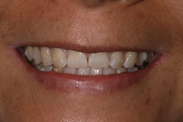
Figure 1
Fig 1. Preoperative, the patient’s existing anterior dentition appeared short proportionally. To esthetically redesign her smile, smile design study was used in determining the ideal proportion, shape, size, color, and style of the restored dentition.
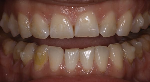
Figure 2
Fig 2. Although the patient had previously completed clear aligner therapy to achieve an improved occlusal scheme, at diagnostic work-up it was evident that the worn dentition required reconstructive treatment to replace severely worn enamel.
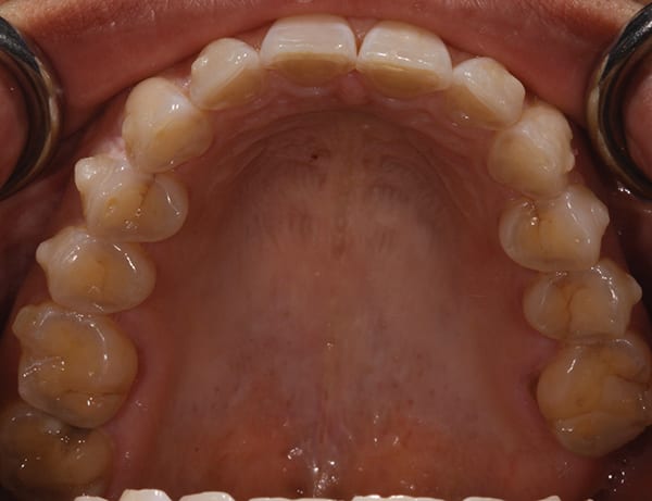
Figure 3
Fig 3. Upon evaluation, it was noted that the patient had worn off all functional guide planes, such as anterior guidance, lateral excursion, and protrusive function.
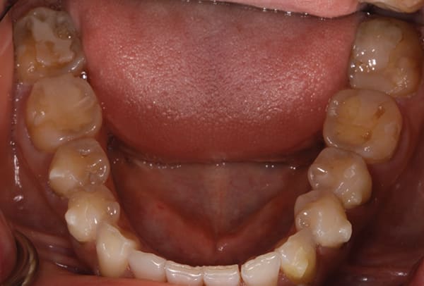
Figure 4
Fig 4. The severe wear required the replacement of missing enamel with a restorative treatment material that would provide optimal strength and occlusal toughness for long-term success.
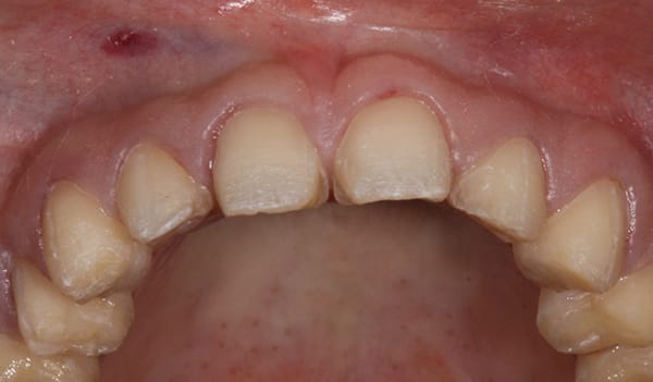
Figure 5
Fig 5. Teeth preparation for both arches (upper arch shown) was performed to remove broken, decayed, and decalcified enamel, leaving a base of healthy enamel.

Figure 6
Fig 6. After final impressions and treatment records were taken, esthetic temporary prototypes were hand-sculpted to develop the patient’s new occlusion and smile design.
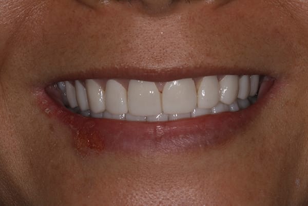
Figure 7
Fig 7. Prior to sending the temporary prototypes for final restoration fabrication, the smile design and occlusal scheme were refined to meet the patient’s esthetic demands and occlusal comfort.
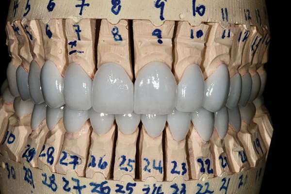
Figure 8
Fig 8. Full-mouth cosmetic and restorative restorations were masterfully sculpted to match the size, proportion, and smile design of the esthetic temporary prototype contours. Following the prototypes eliminated any guesswork.
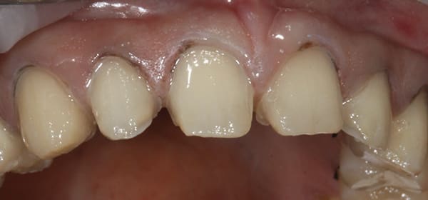
Figure 9
Fig 9. After isolation and decontamination protocols were performed, All-Bond Universal was placed and light-cured for optimal adhesion to promote long-term success.

Figure 10
Fig 10. The anterior porcelain veneers were bonded with Choice 2 luting resin and light-cured. Margins were then refined with a No. 2 scalpel and fine carbide finishing bur.
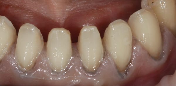
Figure 11
Fig 11. All-Bond Universal adhesive was then placed on the lower anterior teeth to create a hydrophobic surface ideal for long-term success of bonding.
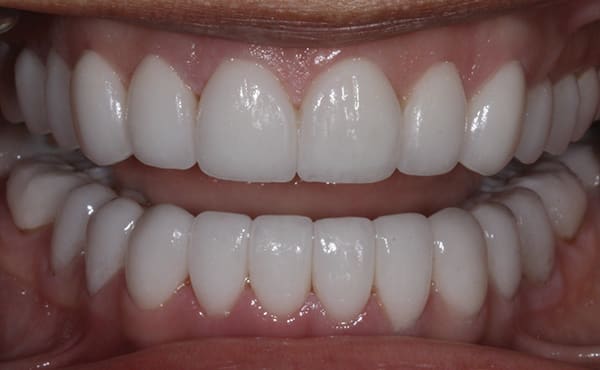
Figure 12
Fig 12. For all molars, full-contour zirconia crowns were bonded using Z-Prime Plus on the intaglio surfaces of the crowns and TheraCem luting resin cement.
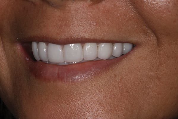
Figure 13
Fig 13. Anterior guidance and canine-protected function was re-established to help ensure long-term success. Optimal tooth position was attained to match the patient’s smile line.
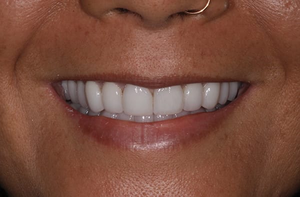
Figure 14
Fig 14. The health, function, and esthetics of the patient’s dentition was restored, and a more youthful smile was achieved.
