In the two cases presented here, Zenostar® zirconia from Wieland Dental (www.wieland-dental.de), a company of the Ivoclar Vivadent Group (www.ivoclarvivadent.com) was used for the final restorations, which were fabricated by Modern Dental Laboratory USA (www.moderndentalusa.com) with support from the Modern Dental USA Digital Processing Center. The restorations were completed using a predictable “textbook” approach plus streamlined communication using digital scans transmitted electronically.
Key Takeaway Points:
• The advantages of the digital workflow are especially apparent with dental implants and other complex cases.
• Using a laboratory that is able to digitally design and manufacture both provisional and final restorations, and which has the technology to scan and transmit traditional impressions, enables improved collaboration, resulting in optimal treatment outcomes.
• A collaborative effort between Modern Dental’s digital team and the authors’ in-house lab and key ceramist led to strong, esthetic restorations that met the patients’ functional and esthetic needs.
Figure Captions
1 & 2. In Case #1, a 59-year-old woman presented with tooth No. 5 fractured at the gumline. Following atraumatic extraction and immediate implant placement, the implant site was scanned chairside (TRIOS® scanner, 3Shape Dental, www.3shape.com) for CAD/CAM design and fabrication, and the laboratory customized a provisional restoration, which was placed by Dr. Clark to maintain/contour tissue during healing. When healing was complete, the site and tissue contours were scanned and electronically transmitted to Modern Dental, where the lab team fabricated the final restoration. A zirconia-to-titanium screw-retained restoration was designed on a 3Shape system, and the facial cutback was designed digitally (Fig. 1). Fig. 2 shows access hole aligned in design.
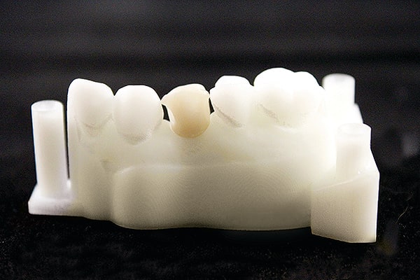
3. After milling, fine-tuning, and sintering, models were finished and cleaned, and the sintered restoration was seated to the titanium interface and fitted to the model. The contacts, occlusion, and contours were verified; no adjustments were required.
4. After the final quality control was performed by the lab, the restoration and printed models were returned to the authors’ office for the porcelain application.
5. The in-house ceramist then layered ceramic over the facial surface of the zirconia substructure for final seating. This allowed for customized esthetics while maintaining the strength of zirconia.
6 & 7. In Case #2, an 80-year-old man presented with tooth No. 15 missing, No. 19 fractured, and a broken crown on No. 18. Tooth No. 19 was atraumatically extracted; immediate implant placement was done at that site and at No. 15. Traditional impressions of all teeth were made and sent to the dental laboratory. A scan abutment was placed on No. 15 and scanned into the 3Shape system, which was used to digitally design a zirconia-to-titanium screw-retained restoration with a facial cutback. After nesting, milling, fine-tuning, and sintering, the restoration (No. 15) was seated to the titanium interface (Fig 6) and fitted to the model (Fig 7). The restoration was then scanned in with the model as the opposing dentition for the design of Nos. 18 and 19.
8. A scan abutment was placed on No. 19, after which teeth Nos. 18 and 19 were designed.
9. No. 18 was designed as a full-contour zirconia with a facial cutback, and No. 19 as a zirconia-to-titanium screw-retained restoration with a facial cutback. Both cutbacks were performed digitally.
10. Final quality control of the case was performed, and all three restorations and the models were carefully packed and returned to the authors’ office, where the in-house ceramist layered facial ceramics for the final seating.
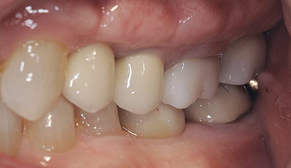
11. Final postoperative photograph.
About the Authors
Wendy AuClair Clark, DDS
Maurice Salama, DMD
Ronald Goldstein, DDS
David Garber, DMD
Private Practice, Atlanta, Georgia


