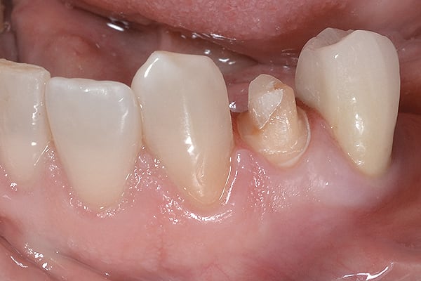
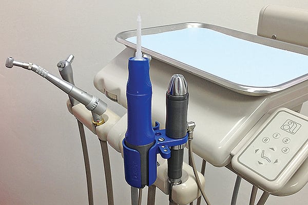
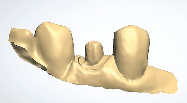
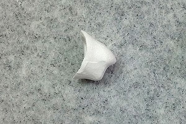
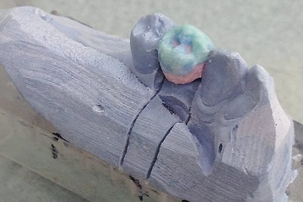
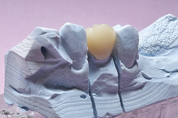
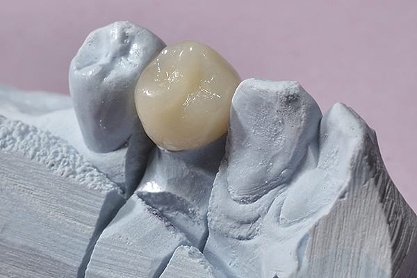
In this case, tooth No. 21 was restored with a layered zirconia crown. The Aquasil Ultra Cordless Tissue Managing Impression System (DENTSPLY Caulk, www.caulk.com) was used to precisely capture the details of the preparation, and the dental laboratory was then able to scan the impression to fabricate a zirconia coping. After the crown was completed, Ceramir® Crown & Bridge (Doxa Dental Inc., www.ceramirus.com) cement was used because of its biocompatibility and ease of handling.
Key Takeaway Points:
Leveraging the strength of zirconia with the added esthetics of layered porcelain can provide beautiful, long-lasting, metal-free restorations.
Aquasil Ultra Cordless Tissue Managing Impression System combines a dispenser for precision placement with a material that is optimized for scanning conventionally recorded impressions.
Partnering with a quality dental lab and selecting an appropriate cement based on substrate and preparation design helps optimize outcomes.
Figure Captions
1. Preoperative radiograph of tooth No. 21. The tooth required full coverage because of a lingual cusp fracture and a large existing mesio-occlusal restoration.
2. Final preparation for full-coverage restoration on tooth No. 21. A layered zirconia crown was selected to provide high strength and esthetics.
3. Aquasil Ultra Cordless digit power™ Dispenser (DENTSPLY Caulk). The Impression Dispenser, with installed intraoral tip, is shown in the blue plastic adapter. On the right is the regulator attached to the dental air line, and at the top of the regulator is a silver knob with settings for flow rate of impression material.
4. Final impression of tooth No. 21 using Aquasil Ultra Cordless Tissue Managing Impression System (DENTSPLY Caulk).
5. Scan of final impression of tooth No. 21, buccal view, using 3Shape™ scanner (www.3shape.com).
6. Scan of final impression of tooth No. 21 using 3Shape™ scanner, showing the planning of the zirconia coping.
7. Milled Cercon® Ht (DENTSPLY DeguDent, www.degudent.com) zirconia coping.
8. Cercon® Kiss (DENTSPLY DeguDent) porcelain layered over zirconia coping, buccal view.
9. Cercon® Kiss porcelain layered over zirconia coping, occlusal view.
10. Final layered zirconia crown on the stone die, buccal view.
11. Final layered zirconia crown on the stone die, lingual view.
12. Internal view of final layered zirconia crown.
13. Completed case after cementation with Ceramir® Crown & Bridge Cement.
14. Final radiograph of tooth No. 21 layered zirconia crown.
Special thanks to Blackburn Dental Laboratory (www.BlackburnDentalLab.com) for the documentation of the crown fabrication process, and the outstanding final result.
About the Author
Nicholas R. Conte, Jr., DMD, MBA
Director of Clinical Research and Education, DENTSPLY Caulk, Milford, Delaware
Clinical Assistant Professor, Rutgers School of Dental Medicine, Newark, New Jersey
Private Practice, Havertown, Pennsylvania

