Abstract
Restoring the beauty of patients’ smiles requires clinicians to incorporate both tooth and gingival esthetics. When gingivectomies and crown-lengthening procedures are required, opting for direct nanohybrid composite restorations can help enable dentists to achieve harmonious blending of the gingival architecture and the anticipated restoration by allowing for tissue healing and subsequent modifications to the gingival portions of restorations. This article presents two cases in which anterior nanohybrid direct composite restorations provided a means to enhance tooth esthetics simultaneous to improving gingival harmony.
Restoring the beauty of patients’ smiles requires clinicians to examine more than tooth structure. A comprehensive smile analysis involves evaluating gingival esthetics, proportion, shade, and other factors in order to be able to propose a treatment plan that will satisfy practitioner and patient expectations.1,2 Among the considerations are bite characteristics and their influence on restoration longevity, pre-existing restorations and the manner in which they can be replaced or altered,3 and financial circumstances that may limit the type of restorative procedure that can be performed.4
In addition, when gingivectomies and crown-lengthening procedures are necessary, an approach is required that enables harmonious blending of the gingival architecture and the anticipated restoration.5 The use of direct nanohybrid composite materials empowers dentists to create esthetic and predictable anterior restorations with the flexibility to accommodate necessary gingival alterations.6 Final tissue healing must be allowed, after which the clinician can smooth and/or add to the gingival portions of restorations.7
Such an approach represents an alternative to traditional crown-lengthening procedures and gingivectomies that occur prior to restorative and/or direct composite placement procedures. As described in Case No. 1, a gingivectomy can be performed first, after which an initial direct composite restoration can be placed, with hemostasis and isolation from blood and crevicular fluids ensured using cotton rolls, retraction cord, and/or rubber dam, as necessary. This facilitates communication with the periodontist about such essential crown-lengthening components as margin placement, shape, and bone level.8,9 Crown lengthening by the periodontist can then be performed approximately 1 week following the initial restorative procedure, with the direct composite restoration being finalized after the tissue has completely healed from the crown-lengthening procedure.
Facilitating this endeavor is the use of nanohybrid composite materials, which demonstrate the esthetic, handling, and material properties required for such cases, eg, Venus Diamond®, Venus Pearl, (Heraeus Kulzer, www.heraeusdentalusa.com). As seen in Case No. 1 with signs of wear, strength is required to withstand masticatory forces; a universal nanohybrid composite (Venus Diamond) that demonstrates enhanced wear resistance, high flexural strength, and low shrinkage stress can be selected.10,11 This nanohybrid composite is formulated with a urethane monomer chemistry that imparts such handling and sculptability properties as firmness and adaptability. These characteristics make shaping, contouring, and polishing composite layers with the incisal cutback technique easier and more efficient.12
When more artistic layering techniques are required, the use of a softer, brushable universal nanohybrid material is an alternative (Venus Pearl). The different particle sizes in the material produce an ideal filler density that contributes to high wear resistance, a non-sticky consistency, and polishability, as demonstrated in Case No. 2.
Case No. 1
A 35-year-old female presented with an implant restoration that had been placed several years earlier for a congenitally missing tooth No. 10. The dentist observed that the restoration, although healthy, did not blend well with the natural dentition and the gingival heights were not harmonious, causing a lack of symmetry. The patient’s chief concerns were finances, lack of symmetry, and the stains and chips on her bonding.
A thorough examination was performed, and no decay was found. The tissue was healthy, but previously placed composite restorations on teeth Nos. 8 and 9 were stained and chipped (Figure 1). The gingival heights on teeth Nos. 7 and 8 were significantly lower than those on teeth Nos. 9 and 10.
By probing sulcus depths and sounding to bone, it was determined that the bone was in a normal relationship to the free gingival margin. This indicated that osseous resection would be required in any area of gingival alteration.13 Osseous modification commensurate to soft-tissue modification was required to provide adequate biologic width.14-16
Clenching, facial pain, and posterior interferences in protrusive position, as well as right and left lateral working and balancing movements, were noted.
Diagnosis
Following the examination, the patient was given a diagnosis of a lack of anterior guidance, unesthetic composite bonding on the anterior maxillary dentition, and unesthetic and unharmonious gingival height symmetry. Therefore, the patient required more gingival length, especially in protrusive guidance, in addition to a change in the shape of her teeth. No other diagnosis was made.
Treatment Planning
Treatment goals for this patient included teeth whitening, creation of a beautiful and symmetrical smile, and establishment of comfortable function by eliminating interferences and pain. This would be accomplished with teeth whitening, followed by equilibration, gingivectomy, and direct composite restorations on teeth Nos. 6 through 11, and crowning lengthening. In addition, impressions would be taken for a bite appliance. Therefore, subsequent to teeth whitening, the treatment plan progressed as follows:
1. Equilibration: Because the patient had posterior interferences over the right and left, equilibration would be addressed prior to initiating composite treatment; the interferences to centric relations were examined, after which composite placement would be performed from cuspid to cuspid. However, prior to removing the posterior excursive interference, the anterior cross-over would be established.17,18
2. Gingivectomy: Next, the periodontist would perform the gingivectomy to move the gingival tissue apically 3 mm based on esthetic parameters. Then, the periodontist would raise the bone 3 mm to provide 3 mm for biologic width.
3. Accurate composite placement: The decision was made to place the composite material prior to the surgical procedure in order to direct the surgeon to the final desired gingival heights. The crown-lengthening procedure on teeth Nos. 7 through 9 and a graft on tooth No. 10 would be scheduled 1 week after restoration placement, with the direct restorations and occlusion finalized upon complete gingival healing.
4. Establishment of incisal guidance: Then, incisal guidance in the cross-over pattern (as determined on an articulator) would be established. A bite analysis was performed using a maxillary trial-smile model (ie, intraoral mock-up) mounted in centric relation with the lower model on an articulator. The trial smile (Figure 2) was created by placing composite directly onto the unprepared teeth, without etching or adhesive, in order to establish an accurate incisal edge position and facial contours. The advantage of an intraoral mock-up compared to a diagnostic waxup is the ability to observe the lower lip position during smiling, speaking, and repose.19 The edges should be just inside the wet zone of the lower lip.
5. Posterior interferences: Then, posterior interferences would be removed.
The trial smile enabled the patient to confirm the proposed changes for her existing direct composite bonding, as well as her desire to alter the shape and length of the implant-supported restoration.20,21 Upon viewing the trial smile and observing for herself the manner in which the proposed restoration for tooth No. 10 now blended more harmoniously with the other three teeth, the patient decided to proceed with receiving four direct composite restorations. In addition, because the patient had financial constraints, she was adamant about completing all restorations in direct composite, not porcelain.
The trial-smile model was then fabricated from an impression of the trial smile, which was also the basis for creating an incisal edge matrix to guide composite placement. The incisal edge matrix, which was created using putty (Flexitime® Putty, Heraeus Kulzer) to illustrate the length and fullness to be added to each tooth, also was made using the trial-smile model.22 After impressions were made, the trial-smile composite was removed and digital photographs were taken.
Clinical Technique
At the restorative appointment, the trial-smile composite was placed over the teeth again, and a permanent marker pen was used to indicate the desired gingival heights (Figure 3). Although the crown-lengthening procedure was planned to be performed by the periodontist 1 week after the restorative appointment, the gingivectomy was done the same day as the direct composite procedure.
The old composite restorations were removed, and no further preparations were necessary, which preserved all the enamel, tooth strength, and integrity. Cotton rolls and cord were placed as necessary to ensure isolation from blood and crevicular fluids. The adhesive bonding protocol was then performed in a two-step process to facilitate the cutback of the incisal portion of the direct restoration.
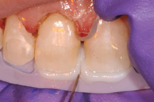
The incisal portion of the teeth was etched (Figure 4), rinsed, and dried, after which a fourth-generation adhesive bonding agent (All-Bond, Bisco, www.bisco.com) was applied and light-cured. The incisal edge matrix was placed in the mouth, and an opacious composite (OB, Venus Diamond) was applied to the incisal edges and light-cured (Figure 5). A cutback of the incisal edge was then completed, with the tips of the cutback reaching into the deepest aspects of the matrix (Figure 6). This technique allowed the more opacious composite to “break up” the areas of translucency in order to duplicate nature.
The entire facial surface was then etched (Figure 7), rinsed, and dried. An adhesive bonding agent was applied and light-cured. Translucent composite was applied to the gingival one-fourth (AM, Venus Diamond) to add chroma and then light-cured. Next, a more opalescent composite (CO, Venus Diamond) was applied to the incisal edges and the entire facial aspect, and then light-cured.
The facial thickness of the composite buildup was verified using a facial contour matrix (Figure 8) that was also based on the trial-smile model and created from putty (Flexitime Putty). This ensured that the restorations would duplicate the same facial thickness observed with the trial smile in the relaxed lip position for facial support.
The porcelain restoration at tooth No.10 was cut back to create space for blending the composite with the adjacent teeth in terms of shape, length, opacity, and shade. The metal substructure was inadvertently exposed, so a pink opaquer (Cosmedent, www.cosmedent.com) was painted over the metal following application of a porcelain etch, silane, and adhesive bonding agent (Figure 9). Then, the same composite shade plan for teeth Nos. 8 and 9 was followed.
The CO composite also was placed on the entire facial aspect of tooth No. 7, as well as the cusp tips of teeth Nos. 6 and 11. After a systematic sequence of finishing and polishing steps,23 the resulting nanohybrid composite restorations demonstrated polychromicity of the individual teeth, in addition to a natural smile appearance.
The appearance of the restorations was enhanced by the crown-lengthening procedure, which was performed by the periodontist 1 week later, to create harmonious gingival heights (Figure 10). The periodontist preferred to wait 1 week following composite placement to perform the procedure to allow slight healing from the gingivectomy. Waiting too long would have resulted in tissue inflammation due to lack of biologic width.
Case No. 2
A 16-year-old female presented with discolored and chipped composite restorations on teeth Nos. 8 and 9; tooth No. 9 appeared dark due to nonvitality (Figure 11). Her chief concerns were the stained and chipped composite restorations. The examination revealed unharmonious gingival heights and incisal edges that did not follow the curve of the lower lip. The patient’s tissue was healthy, and the examination showed no decay or occlusal issues.
The sulcus measured 3 mm without anesthesia to the natural attachment. Because bone had not been reached, it would be possible to perform a gingivectomy and remove 1.5 mm of gingival tissue while still leaving 1.5 mm of the patient’s natural sulcus.16
Therefore, treatment goals included correcting the smile line to create harmony between the incisal edges and the lower lip, as well as creating a harmonious gingival display (ie, relationship of gingival heights and upper lip). In addition, the dark tooth No. 9 would be blended with the natural dentition.
Treatment Planning
A trial smile and a bite analysis were performed, and an incisal matrix was made, as described in Case No. 1. A gingivectomy would be performed to remove 1.5 mm of gingival tissue, leaving 1.5 mm of the patient’s natural sulcus. Nanohybrid composite restorations would be placed on teeth Nos. 6 through 10. The composite restorations would be finalized after gingival healing.
The negative space between the incisal edges and lower lip would allow a thicker composite layer in that area. This would provide the clinician with more room for blocking out the underlying darker color on tooth No. 9.24 In addition, the added fullness and length would give the patient a greater presence in her smile.
Teeth Nos. 7 through 10 would be restored following the same two-stage technique described in Case No. 1. This two-stage process is necessary when clinicians use the cutback technique to create subtle patterns in the incisal translucency.25 Only the cusp tip of tooth No. 6 would be bonded to create equal length with tooth No. 11, as well as serve as protection for the incisal edges of teeth Nos. 7 and 8.
Clinical Technique
Crown lengthening was not necessary in this case due to the excessive sulcus depths (ie, 3 mm).16 The initial sulcus depths were greater than 3 mm, and 1.5 mm of gingival tissue were removed to create a harmonious gingival line while still maintaining 1.5 mm of the patient’s natural sulcus. The gingivectomy was performed the same day as the direct composite placement procedure.
Following the same adhesive and layering sequence described in Case No. 1, an opacious nanohybrid composite (OB, Venus Pearl) was placed on the incisal edges prior to the cutback to create a subtle translucency pattern. The incisal matrix was used to verify the cutback on tooth No. 9 and prior to rinsing and cleaning the area in preparation for the second bonding stage (Figure 12).
After the cutback, a more translucent composite (CO, Venus Pearl) was applied to the entire facial surface, with a B1 shade placed on the gingival one-fourth to create natural polychromicity. A fine polishing point (Hereaus) was used to remove any interproximal scratches (Figure 13). The smoothness of the final composite restorations would prevent staining and enable the patient to clean more effectively (Figure 14).
Conclusions
The case presented in this article demonstrates the manner in which anterior nanohybrid direct composite restorations can be provided a to patient in order to best meet his/her needs and expectations. These included refining the gingival portion of the smile, as well as preserving natural tooth structures. Although direct composite restorations do require maintenance, choosing this minimally invasive option leaves a healthy foundation on which to place permanent porcelain restorations in the future, if desired.
About the Author
Susan Hollar, DDS
Private Practice
Arlington, Texas
References
1. Frese C, Staehle HJ, Wolff D. The assessment of dentofacial esthetics in restorative dentistry: a review of the literature. J Am Dent Assoc. 2012;143(5):461-466.
2. Bitter RN. The periodontal factor in esthetic smile design—altering gingival display. Gen Dent. 2007;55(7):616-622.
3. Donitza A. Creating the perfect smile: prosthetic considerations and procedures for optimal dentofacial esthetics. J Calif Dent Assoc. 2008;36(5):335-340, 342.
4. Feigenbaum N. The challenge of cost restrictions in smile design. Pract Periodontics Aesthet Dent. 1991;3(6):41-44.
5. Robbins JW. Direct composite bonding in conjunction with surgical tissue management. Oper Dent. 2004;29(3):347-349.
6. Portalier L. Diagnostic use of composite in anterior aesthetics. Pract Periodontics Aesthet Dent. 1996;8(7):643-652.
7. Hempton TJ, Dominici JT. Contemporary crown-lengthening therapy: a review. J Am Dent Assoc. 2010;141(6):647-655.
8. Certosimo FJ, Connelly ER, Paul BF, Klier KR, Fitch DR. Accessing restoration margins—a multidisciplinary approach. Gen Dent. 2000;48(3):278-282.
9. Block PL. Restorative margins and periodontal health: a new look at an old perspective. J Prosthet Dent. 1987;57(6):683-689.
10. Koottathape N, Takahashi H, Iwasaki N, Kanehira M, Finger WJ. Two- and three-body wear of composite resins. Dent Mater. 2012;28(12):1261-1270.
11. Marchesi G, Breschi L, Antoniolli F, Di Lenarda R, Ferracane J, Cadenaro M. Contraction stress of low-shrinkage composite materials assessed with different testing systems. Dent Mater. 2010;26(10):947-953.
12. Venus Diamond and Venus Diamond Flow. [Brochure]. South Bend, IN: Heraeus, 2010.
13. Cunliffe J, Grey N. Crown lengthening surgery—indications and techniques. Dent Update. 2008;35(1):29-30, 32, 34-5.
14. Kois JC. The restorative-periodontal interface: biologic parameters. Periodontol 2000. 1996;11:29-38.
15. Ganji KK, Patil VA, John J. A comparative evaluation for biologic width following surgical crown lengthening using gingivectomy and ostectomy procedure. Int J Dent. 2012;2012:479241.
16. Spear F. Using margin placement to achieve the best anterior restorative esthetics. J Am Dent Assoc. 2009;140(7):920-926.
17. FitzGerald LJ. Restoring anterior guidance by use of composite resin. Cranio. 1996;14(3):182-185.
18. Solow RA. Equilibration of a progressive anterior open occlusal relationship: a clinical report. Cranio. 2005;23(3):229-238.
19. Kovacs BO, Mehta SB, Banerji S, Millar BJ. Aesthetic smile evaluation—a non-invasive solution. Dent Update. 2011;38(7):452-454, 456-458.
20. Pulliam RP, Green S. Collaborative care for better aesthetic outcomes. Dent Today. 2011;30(11):152-154.
21. Adar P. Avoiding patient disappointment with trial veneer utilization. J Esthet Dent. 1997;9(6):277-284.
22. LeSage BP. Aesthetic anterior composite restorations: a guide to direct placement. Dent Clin North Am. 2007;51(2):359-378, viii.
23. Glazer HS. Simplifying finishing and polishing techniques for direct composite restorations. Dent Today. 2009;28(1):122, 124-125.
24. Jarad FD, Griffiths CE, Jaffri M, Adeyemi AA, Youngson CC. The effect of bleaching, varying the shade or thickness of composite veneers on final colour: an in vitro study. J Dent. 2008;36(7):554-549.
25. Villarroel M, Fahl N, De Sousa AM, De Oliveira OB Jr. Direct esthetic restorations based on translucency and opacity of composite resins. J Esthet Restor Dent. 2011;23(2):73-87.
Go online to read a second case exemplifying this technique. dentalaegis.com/go/cced430


