Patients often present with dental problems that require a team approach in which the restorative dentist works with other specialists to offer the patient the best outcome. In fact, the “team approach” led to the author publishing the first interdisciplinary textbook devoted to esthetic dentistry, Esthetics in Dentistry, in 1976.1 Over the past 55 years, the author has also collaborated with numerous trusted specialists, including current practice partners, orthodontist and periodontist Maurice Salama, DMD, and David Garber, DMD, who is dual trained in prosthodontics and periodontics. Additional associates consist of Henry Salama, DMD, a dual degree periodontist/prosthodontist; two prosthodontists, Maha El- Sayed, DMD, and Wendy Clark, DDS, MS; oral surgeon Abtin Shahriari, DMD; and another general dentist, Nadia Esfandiari, DMD. The author still practices today as he did 50 years ago; collaborative consultations typically took place in either his general dentist’s office or by referring the patient to different specialists. Although it took many years to create the team of specialists, the group consultations now take place in the office, which was built to easily a commodate interdisciplinary treatment planning. Nevertheless, thanks to new technology, collaborators from around the country or around the world can communicate digitally, remotely by telephone, e-mail, with Skype or videoconferencing. Therefore, it is even more feasible for the general dentist to refer to different specialist offices wherever they may be.
The author has long been a proponent of the team approach to patient care in situations such as those illustrated in the cases discussed in this article. He has always taught that “esthetic dentistry” includes virtually all phases of dentistry, from preventive dentistry to endodontics, orthodontics, oral surgery, periodontics, prosthodontics, psychology, pedodontics, and everything in-between. However, he considers it essential that, as the restorative dentist, he oversees the care, acting as the “captain” of the team, being careful to first complete a number of steps before summoning colleagues who may ultimately be involved in treatment. These steps include the clinical examination, digital photographs, radiographs, periodontal charting, occlusal analysis, diagnostic models, and esthetic analysis. Another essential factor in the interdisciplinary practice is having a well-trained staff that needs to be educated on the role of the different specialties as they relate to specific patient problems.2,3
Intraoral Examination
The author believes it is most important, however, to start with a tooth-by-tooth intraoral examination. This is something every patient should receive, and it is the basis of the initial consultation with the patient. This exam is also an excellent way to build trust based on an understanding of the patient’s presenting dental condition. It is also the best way for dentists to document their findings for legal purposes should there ever be a question about what and why treatment was performed.
Unless the dentist takes the photographs himself—as the author does in about 20% of cases—it is best to wait to meet with the patient until the trained dental assistant takes all necessary digital photos and they are available on a chairside monitor. Doing so allows the dentist to review and present this objective information chairside showing the existing condition of each tooth at presentation. With patients being informed at the onset of their condition, they are less likely to blame the new dentist for pain or other problems related to preexisting conditions such as cracks and/or decay revealed upon removal of a restoration. Patients need to see the dentist as the solution, not the problem.
If a dentist does not take photographs or intraoral images, there will be no record of the patient’s actual condition. Simply placing a note in the patient’s folder stating that there is deep decay is not sufficient. The picture is a visual reminder for the patient, both then and later. Even when the patient does not take the dentist’s advice, such as a crown or veneer when translumination reveals microcracks in the enamel, there is a proper photographic record of what was present. Therefore, if a problem occurs later the dentist has an appropriate record of his previous advice.
The author considers cavity detection devices such as DIAGNOdent (KaVo Dental, www.kavousa.com) and CarieScan™ (CarieScan Ltd, www.cariescan.com) indispensable aids in treatment planning because they can reveal potential cavities or decay in the mouth, demonstrating the need to lower the bacterial content to ensure that restorations will last longer. In addition, both the Carestream CS 1600 (Carestream Dental, www.carestreamdental.com) and the Soprolife (Acteon Group, www.acteongroup.com) intraoral cameras are beneficial to use before proceeding with restorations because they can disclose caries—a colored light enables the clinician to easily determine if all decay is removed before any restorative material is applied.
Communication with Specialists
Usually, the only consultation needed by a general restorative dentist is with the laboratory, but, depending on the situation, consultation may be necessary with periodontists, orthodontists, oral surgeons, endodontists, or even plastic surgeons. Each patient should be evaluated from the standpoint of improvements that can be made considering each of dentistry’s specialty areas. This is where consultations come into play in a treatment plan managed by the restorative dentist, who remains the central decision-maker to ensure that the patient is continuing in the planned steps. Again, the new communication tools make it much more feasible for the specialist to continue to inform the general dentist via digital photographs throughout treatment in “real-time.” This makes it possible for such consultations to take place either onsite or remotely. Additionally, whatever means are used, it is best to have continuous coordinated documentation for the patient who has interdisciplinary problems.
Patient Management Tips
One of the toughest challenges the general practitioner faces is forecasting which patients might be especially difficult to treat. To minimize the chance of having a dissatisfied patient, there are certain techniques the author finds helpful.
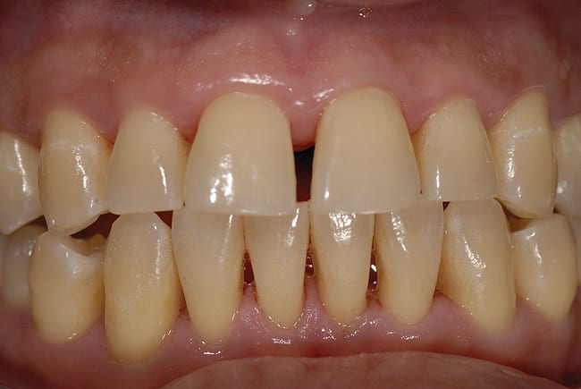
Dealing with a patient who has a specific request may require an approach that does not directly address the request. Because many patients may not actually know what they want, simply agreeing to a specific request may not meet the patient’s real desire. For example, when a patient once asked to have her crowns changed to porcelain to improve her appearance, the author responded with a question: “If I had a magic wand, and could wave it across your face, is this the smile you would have—just four new porcelain crowns?” Upon learning that what the patient really wanted was a “beautiful smile with white teeth,” the author instead suggested that he be allowed to design a treatment plan with that objective. Naturally, the patient consented and an interdisciplinary plan was formulated. (See Case 1.)
Also, to clarify patient expectations and inform specialists of the goals of treatment, the author recommends offering a preview of results with either or both esthetic imaging and/or a “trial smile,” which can be done using, for example, a vacuform matrix/or siltek with composite resin. This enables the patient to “try on” the smile for a period before having the final treatment performed. Its main purpose is to prevent failure and to enable patients to understand what can and cannot be accomplished. Digital photos must be taken so the patient can see him or herself in two dimensions as well as three dimensions such as looking into the mirror.
Then there are bona fide “difficult patients,” those who are body-dysmorphic or inclined to focus on a tooth problem rather than their bigger issues in life such as their social life, marriage, business, family, financial problems, or inadequacies within themselves. Some dentists may be tempted to take on such difficult patients for economic reasons—especially in a down economy—but it is important for dentists to be able to identify patients they can treat successfully versus those they may have difficulties with. Plainly put, there are certain patients whom a dentist simply cannot please. The author’s How to Manage Difficult Patients Before They Manage You course presents 55 years of different classifications of patients and how they were successfully managed, giving tips to the full team in each situation.
Specialists such as orthodontists, periodontists, and others can be indispensable in such situations because they are able to see the patient from a different perspective. The author urges heeding the advice of a specialist or even a staff member who warns that a particular patient will be difficult to please.4
In addition to documentation, including extraoral and intraoral images, as well as a trial smile to enable patients to preview treatment plan goals, patient consent is paramount, especially when treatment involves compromises.” The patient must sign a written document, and it must be well documented in the patient’s folder that all treatment options were presented even though the patient declined ideal treatment suggestions. Patients tend to forget that the dentist verbally suggested ideal specialty treatment; this is the main reason why the assistant must make certain that all treatment suggestions are clearly documented in the patient’s folder, especially when the patient elects a compromised treatment plan.
Team-Treated Patient Cases
The author presents a sampling of his cases to illustrate interdisciplinary treatment plans.
Case 1
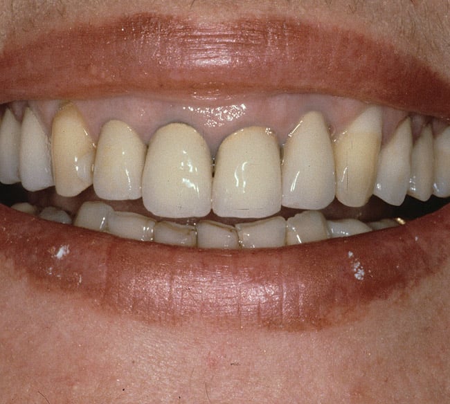
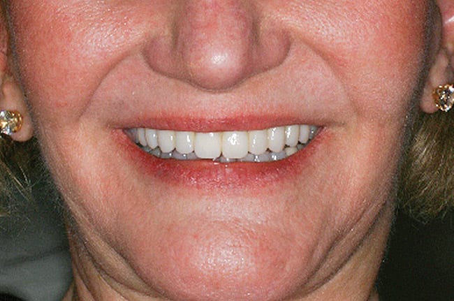
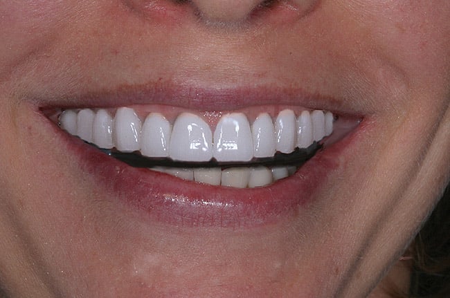
To address this patient’s desire of attaining a beautiful smile with white teeth, the treatment plan went far beyond her request of changing just her four anterior porcelain crowns. The patient had relatively good bone, but erosion on the bottom incisal edges, chipped and fractured teeth, bad fillings, as well as a high lip line were all problems that needed to be addressed (Figure 1). After considering the diagnostic factors, it was determined that the patient’s high lip line should, if possible, be made into a medium lip line by crown lengthening periodontal surgery. In presenting the treatment plan, it was important that the patient see the plan as the clinician did through esthetic imaging and diagnostic wax-ups. The conservative plan entailed bleaching the lower teeth, crown lengthening, cosmetic contouring, bonding the lower anteriors, crowning, and four porcelain veneers. The patient was able to have all of the recommended treatment done and was highly satisfied with the result (Figure 2). This represents an example of a patient who presented with a certain treatment in mind—porcelain crowns—but who was amenable to the author’s professional advice.
Case 2
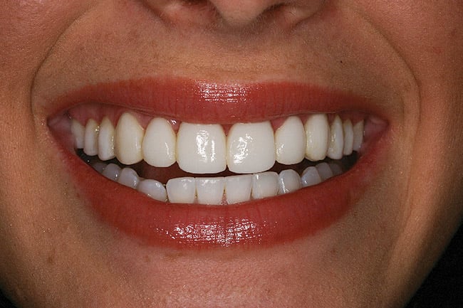
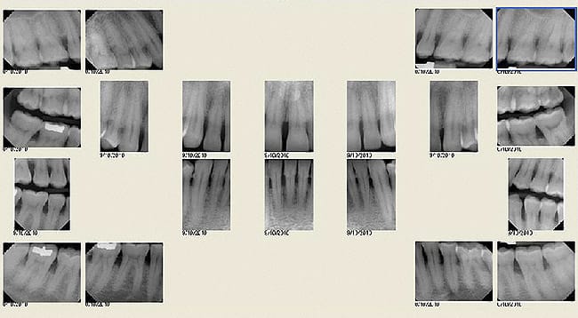
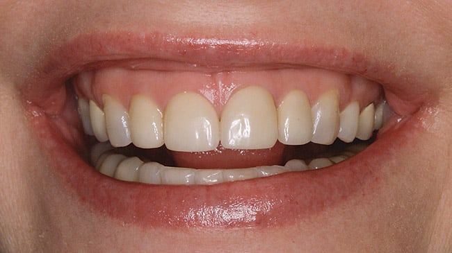
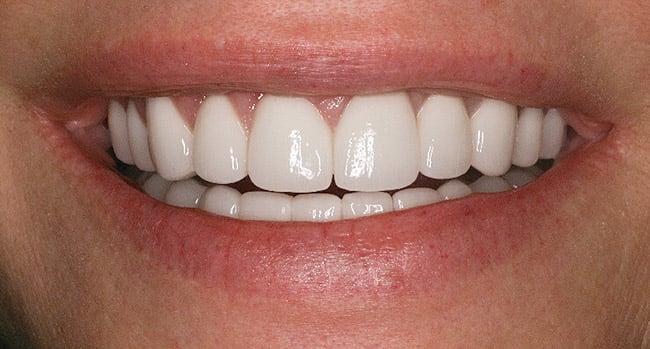
This is an example of a patient who was quite difficult to please (Figure 3). She presented with advanced bone loss (Figure 4) and had been told by multiple dentists, including both general practitioners and specialists, that she needed to have her teeth extracted. Because she was adamantly opposed to extraction, the author suggested a “three-phase fix,” working first with a fixed temporary splint, then a periodontist, and, finally, proceeding to the restorative phase. Offering no guarantees of longevity, the author agreed to try full-arch temporary splinting for a period of time to ensure a satisfactory functional response, before creating the final restoration. However, despite her initial desire to save her teeth, the patient totally shifted her focus to her appearance, which was significantly improved by the temporary restoration. Instead of having the treatment completed by the author, she had the final treatment done by another dentist, only to return a year later with complaints related to breakage, caries, and margins that did not fit. After the author resumed the case to create the final restorations, the patient continued to bring in new pictures of faces much younger than hers to show how she wanted to look. The final result eventually pleased her (Figure 5 and Figure 6), but it took her husband to verify she had achieved the best that dentistry could accomplish both esthetically and functionally before she was satisfied.
Case 3
The treatment plan for this young woman who was unhappy with her smile combined cosmetic periodontal surgery, bonding, porcelain veneers, and all-ceramic crowns. Although she was initially pleased with what the previous dentist had accomplished, she became dissatisfied with the tooth proportions, especially the width of the teeth (Figure 7 and Figure 8). The author suggested crown lengthening and, for economic considerations, direct composite resin bonding the posteriors—a total of two porcelain veneers and four Procera® (Nobel Biocare, www.nobelbiocare.com) crowns. He also explained to the patient that a new hairstyle would make her face look less wide. Utilizing a team approach, with Dr. Garber doing the crown lengthening, once again collaboration and interdisciplinary dentistry made it possible to achieve the results pleasing to the patient, her family, and the restorative dentists (Figure 9 and Figure 10).
Case 4
This patient, an Italian runway model, wanted to improve her smile in a short period of time to fulfill a lucrative photographic modeling contract—giving the author less than 6 weeks to complete the treatment. The case began with immediate crown-raising periodontal surgery by Dr. Salama, plus a removable appliance to improve the position of her teeth, and, finally, porcelain veneers on the maxillary arch. Photographs show her prior to veneer placement (Figure 11), then after, with good inter-incisal distance achieved (Figure 12). The author believes a more natural look can be achieved by opening up the incisal embrasures. Otherwise, the dentition appears too joined, giving a “false” look.
Case 5
This case demonstrates the importance of obtaining accurate restoration margins. The patient had 10 porcelain veneers fabricated in another country, and she complained of gingival pain and tooth sensitivity. The clinical examination revealed the veneer margins were fitting 1 mm to 1.5 mm away from the tooth (Figure 13). In fact, none of the veneers fit properly, exudate was coming from most gingival areas, and all the veneers required initial reshaping. While the patient wanted the veneers removed, the author explained that it was first necessary to recontour the gingival margins and allow the tissue to heal. The porcelain was then polished to make it smooth. Nevertheless, the patient later wanted to have the veneers properly remade.
Case 6
Saving the papilla is one of the restorative dentist’s biggest concerns, because no matter how a tooth is shaped, the papilla affects the tooth form. To avoid “black triangles,” a conservative periodontic approach involves the use of laser-assisted new attachment procedure (LANAP). Although the patient had advanced bone loss (Figure 14 through Figure 16), there was absence of pain 24 hours following LANAP procedure performed by Dr. Salama. Four months healing showed new bone attachment (Figure 17) and absence of interdental tissue loss (Figure 18).
Case 7
This patient was concerned with her aging worn and discolored smile (Figure 19 and Figure 20). The treatment plan started with a trial smile, with esthetic imaging to enable the patient to visualize why the recommended treatment would involve lengthening the teeth, raising the gum tissue, and building out the buccal corridor (Figure 21 and Figure 22). Following crown lengthening, 12 Procera all-ceramic maxillary crowns were constructed. Conservative bleaching and cosmetic contouring was performed on the mandibular arch. All-ceramic crowns were chosen due to the patient’s multiple large restorations and previous bruxism habit. A full nightguard was constructed following the final crown seating (Figure 23), which consisted of soft inside and hard acrylic outside.
Case 8
This 22-year-old waitress was too embarrassed to smile (Figure 24), which limited her full potential both socially and in reaching her career goals. Porcelain veneers and a resin-bonded fixed all-ceramic bridge were made without reducing the tooth structure. (This can best be accomplished when the teeth need building out or when multiple spaces exist.) The result (Figure 25 and Figure 26) was a life-changing physical transformation for this young woman, who after her smile makeover went on to earn a degree in finance.
Case 9
In this case, the author and his orthodontist partner, Dr. Salama, chose to redo a case that did not meet their own standards. To correct an arch irregularity and improve the lip line (Figure 27 and Figure 28), the plan was to contour the teeth, then proceed with crown lengthening followed by all-ceramic crowns on the maxillary arch and all-ceramic crowns and six porcelain veneers on the lower arch. Although the result was satisfactory to the patient, it did not meet the practitioners’ standards, so the surgical treatment was redone with new all-ceramic crowns on the maxillary arch. The final result then pleased both of the doctors and the patient (Figure 29 and Figure 30).
The Future of Dentistry
The author sees a future in which full-body imaging will include the oral cavity, including teeth, and artificial intelligence-generated treatment plans will use holographology for diagnosis, treatment, instruction, and study. Dentistry will also use stronger materials and more creative methods, and science will offer robust genetically engineered teeth, among many other positive strides. Eventually, each treatment room will have voice-activated recording devices enabling extremely accurate documentation to record what both dentist and patient discussed and agreed to. Finally, in the next decade dentistry will see the increasing influence of general dentists developing closer team working relationships to the benefit of all patients.5-7
About the Author
Ronald E. Goldstein, DDS
Clinical Professor of Oral Rehabilitation,
Georgia Health Sciences Universit
Augusta, Georgia
Adjunct Clinical Professor of Prosthodontics,
Boston University Henry M. Goldman School of Dental Medicine
Boston, Massachusetts;
Adjunct Professor of Restorative Dentistry
The University of Texas Health Science Center
San Antonio, Texas
Private Practic
Atlanta, Georgia
References
1. Goldstein RE. Esthetics In Dentistry. Philadelphia, PA: J.B. Lippincott; 1998:17-49.
2. Goldstein R. Cosmetics expert Dr. Ron Goldstein shows how working together works. Interview by Cathy Jameson. Dent Teamwork. 1993;6(2):18-23.
3. Levin RP. Why Most Practices Don’t Have a Dream Team. Dentistry Today Web site. March 24, 2011. https://www.dentistrytoday.com/management-articles/4798-why-most-practices-dont-have-a-dream-team. Accessed August 30, 2012.
4. Golden RE. Treating Difficult and Challenging Patients (Part 1). Contemporary Esthetics and Restorative Practice. 2003;7(5):20-23.
5. Spear FM, Kokich VG. A multidisciplinary approach to esthetic dentistry. Dent Clin North Am. 2007;51(2):487-505.
6. Spear FM. Forming an interdisciplinary team: a key element in practicing with confidence and efficiency. J Am Dent Assoc. 2005;136(10):1463-1464.
7. Chiche GJ, Fahl N Jr, Kois JC. The changing world of dentistry. Dent Today. 2011;30(2):126,130,132-133.
